
Received 2025-04-21
Revised 2025-08-02
Accepted 2026-02-10
Investigating the Effect of Metformin on p53 Protein Using a Molecular Docking Approach
Short title: Investigating the Effect of Metformin on p53 Protein Using a Molecular Docking Approach
Farshid Aaliazar 1, Bahare Shadazfa 2, Mohammad Parsafar 3, Mohammad Ali Rahmani 4
1 Department of Medical Genetics, Technofest Institute of Technology (TITU), Belgium, Erquelinnes
2 Department of Cellular and Molecular Biology, Tehran Islamic Azad University of Medical Sciences, Tehran, Iran
3 Department of Chemistry, Faculty of Science, Iran University of Science and Technology, Tehran, Iran
4 Department of Surgery, Yasuj University of Medical Sciences, Yasuj, Iran
|
Abstract Background: The tumor suppressor protein p53 plays a fundamental role in maintaining genomic stability by regulating the cell cycle, DNA repair, and apoptosis. Metformin, a widely prescribed antidiabetic drug, has demonstrated potential anticancer properties, but the exact molecular mechanisms underlying its effects remain fully elucidated. This study explored the molecular interaction between metformin and the p53 protein using a computational molecular docking approach to understand better how metformin may influence p53’s tumor-suppressive functions. Materials and Methods: The crystal structure of human p53 (PDB ID: 1KZY) was prepared using UCSF Chimera, and metformin’s structure was optimized via OpenBabel and Chimera. Binding site prediction was performed using DeepSite, and molecular docking simulations were conducted with AutoDock Vina in PyRx. Interaction analyses were performed in UCSF Chimera, PyMOL, and PDBsum to identify hydrogen bonds, hydrophobic interactions, and electrostatic interactions. Results: The docking analysis indicated that metformin binds to p53 with a high binding affinity of −8.6 kcal/mol. Hydrogen bonds with Ar273 (2.5 Å) and Glu285 (2.8 Å), hydrophobic interactions with Asp281, Val272, and Phe270, and electrostatic interactions with Lys120. Nine binding poses were analyzed, suggesting that metformin may bind flexibly to the p53 binding pocket. The residues involved are critical for DNA binding and structural stability, indicating that metformin could contribute to the stabilization and functional regulation of p53. Conclusion: This study provides computational evidence that metformin directly interacts with important functional residues of the p53 protein, supporting the hypothesis that it may enhance p53’s tumor-suppressive activity. These findings offer a molecular basis for the anticancer effects of metformin and highlight the need for further in vitro and in vivo studies to confirm and expand upon them, especially in cancers with p53-related dysregulation. [GMJ.2026;15:e3872] DOI:3872 Keywords: P53; Metformin; Molecular Docking; Protein–ligand Interaction; Cancer Therapy |
|
GMJ Copyright© 2026, Galen Medical Journal. This is an open-access article distributed under the terms of the Creative Commons Attribution 4.0 International License (http://creativecommons.org/licenses/by/4.0/) Email:gmj@salviapub.com |

|
Correspondence to: Mohammad Ali Rahmani, Department of Surgery, Yasuj University of Medical Sciences, Yasuj, Iran. Telephone Number: +987132305410 Email Address: mohammadali.rahmani92@gmail.com |
|
GMJ.2026;15:e3872 |
www.salviapub.com
|
Aaliazar F, et al |
Investigating the Effect of Metformin on p53 Protein Using a Molecular Docking Approach |
|
2 |
GMJ.2026;15:e3872 www.salviapub.com |
Introduction
The p53 protein, often called the “guardian of the genome,” is a critical tumor suppressor that regulates the cell cycle, DNA repair, and apoptosis. The TP53 gene encodes it and is one of the most frequently mutated genes in human cancers. Under normal conditions, p53 maintains genomic stability by inducing cell cycle arrest or apoptosis in response to cellular stress, such as DNA damage or oncogene activation. However, dysfunction of p53, either through mutation or inactivation, is associated with tumor progression, resistance to therapy, and poor prognosis in various cancers [1].
Metformin, a biguanide derivative, is widely prescribed as a first-line treatment for type 2 diabetes due to its efficacy in lowering blood glucose levels, it also exhibits potential anticancer properties. Emerging evidence suggests that metformin may also interact with p53, potentially enhancing its tumor-suppressive functions. For instance, metformin has been shown to activate p53 in cancer cells, leading to the induction of apoptosis and inhibition of tumor growth [2]. Although numerous studies have explored the anticancer effects of metformin, most have focused on its indirect modulation of cellular signaling pathways, such as AMP-activated protein kinase (AMPK)-mTOR inhibition of mitochondrial complex I, and IGF-1, without providing detailed structural insights into a direct interaction with p53 [2, 3]. To date, the precise binding site of metformin on p53 and the specific residues involved in this potential interaction have not been clearly identified [4]. Moreover, high-resolution three-dimensional visualizations of metformin–p53 complexes are scarce, and few studies have employed complementary visualization platforms, such as UCSF Chimera and PyMOL, to illustrate docking orientations and key molecular contacts. The integration of molecular docking predictions with the known functional domains of p53, particularly those critical for DNA binding and structural stability, remains limited, leaving a gap in linking computational results to biological significance. Furthermore, there is a lack of testable structural hypotheses derived from docking models that could be experimentally validated through site-directed mutagenesis, binding assays, and functional analyses, which are essential steps toward confirming and expanding the understanding of metformin’s potential role in modulating p53 activity.
Despite promising findings, the precise molecular mechanisms underlying the interaction between metformin and p53 remain poorly understood. Molecular docking is a powerful computational tool that predicts the binding affinity and interaction patterns between small molecules and target proteins. By simulating the binding process, docking research offers valuable knowledge about the structural basis of ligand-protein interactions, which can guide drug discovery and development [5-7]. In this study, we employed molecular docking using PyRx and Chimera to investigate metformin binding to the p53 protein. The primary objective of this research was to elucidate the binding affinity and interaction patterns of metformin with p53 and shed light on the potential mechanisms by which metformin modulates p53 activity.
Materials and Methods
Protein Preparation
The crystal structure of human p53 (PDB ID: 1KZY) was retrieved from the Protein Data Bank (available at www.rcsb.org). Pre-processing was carried out in UCSF Chimera (version 1.19), where crystallographic water molecules and non-essential heteroatoms were removed. Polar hydrogens were added, and protonation states of ionizable residues were assigned to reflect physiological pH (7.4). Energy minimization was performed using the AMBER ff14SB force field with steepest descent and conjugate gradient steps to relieve steric clashes and optimize geometry. The final minimized structure was saved in PDB format for docking [8, 9].
Ligand Preparation
The 3D structure of metformin was obtained from the PubChem database (CID: 4091) in SDF format and converted to PDB format using OpenBabel (available at www.cheminfo.org/Chemistry/Cheminformatics/FormatConverter/index.html). Geometry optimization and energy minimization were performed in Chimera using the GAFF force field to generate the lowest-energy conformation. Gasteiger charges were assigned, and rotatable bonds were defined before saving the ligand in PDBQT format for docking simulations [10, 11].
Binding Site Prediction
Potential ligand-binding pockets of the p53 protein were predicted using DeepSite, a convolutional neural network–based web server (available at www.playmolecule.org). The pre-processed p53 structure (PDB ID: 1KZY) was uploaded to the server, and predicted pockets were ranked according to their calculated probability scores. The highest-ranked pocket, corresponding to the site with the greatest predicted ligand-binding propensity, was selected as the docking target for subsequent molecular docking analyses [12].
Molecular Docking
Molecular docking simulations were conducted using AutoDock Vina integrated into the PyRx virtual screening platform. The grid box dimensions were set to 33.45 × 52.30 × 45.25 Å and centered on the binding site predicted by DeepSite, ensuring comprehensive sampling of potential ligand orientations and conformations. All other docking parameters were kept at default values, except the exhaustiveness, which was set to 8 to improve search accuracy. For validation of the docking protocol, the co-crystallized ligand (when available) was re-docked into its original binding site, and the resulting pose was compared with the experimentally determined conformation to assess the reliability of the docking methodology [13].
Binding Affinity and Interaction Analysis
The binding affinity (ΔG, kcal/mol) for the metformin–p53 complex was obtained directly from the scoring function implemented in AutoDock Vina. The top-ranked docking pose was selected for further examination based on its lowest predicted binding free energy. Visualization and interaction analysis were performed using UCSF Chimera and PyMOL (version 3.1.1). Hydrogen bonds were identified by measuring donor–acceptor distances ≤3.6 Å and angles ≥120°, while hydrophobic contacts were defined by carbon–carbon distances ≤4.5 Å. Electrostatic interactions were assessed by evaluating proximities between oppositely charged side chains and ligand functional groups. Interacting residues within 4 Å of metformin were cataloged, and all key contacts were annotated in the three-dimensional visualizations prepared for publication-quality figures [5, 7].
Results
Binding site prediction through DeepSite detected potential ligand-binding pockets in the p53 protein. The highest-ranked site within the DNA-binding domain was considered in docking simulations. Molecular docking of metformin into this pocket with AutoDock Vina resulted in a top-ranked pose with predicted binding energy (ΔG) of −8.6 kcal/mol, indicating a thermodynamically stable interaction. Nine docking results were obtained, with binding energies ranging from -8.6 kcal/mol to -0.6 kcal/mol (Table-1).
Following interaction analysis between the receptor and the ligand through the PDBsum, it was determined the type of interaction, the distance between the protein residue and metformin, and the importance of each residue in the interaction (Table-2). Arg273 forms a hydrogen bond with metformin, stabilizing the complex. Accordingly, Asp281 is involved in hydrophobic interactions and contributes to structural stability. These findings indicate that metformin specifically interacts with some residues of the p53 protein and potentially affects its activity.
Three-dimensional visualization of the docked complexes in UCSF Chimera and PyMOL revealed deep positioning of metformin within the binding pocket, forming some stabilized interactions with key residues (Figure-1). In the binding site analysis, all interacting residues were within 4 Å of the ligand, supporting the predicted high-affinity binding poses. Two hydrogen bonds were observed between metformin and the side chains of Arg273 and Glu285, with donor–acceptor distances ranging from 2.5 to 2.9 Å. A further three hydrophobic interactions were observed with Asp281, Val272, and Phe270, as well as an electrostatic interaction with Lys120. Residues in this functional region of p53 are critical for DNA binding and structural stability. The consistency of interactions across different visualization platforms, combined with the short hydrogen bond distances, validates the high-affinity binding poses predicted (ΔG = −8.6 kcal/mol) and suggests that metformin may induce the stabilization of p53’s active conformation, thereby enhancing its tumor-suppressive effect.
Discussion
In this study, molecular docking analyses demonstrated a strong and favorable interaction between metformin and the p53 tumor suppressor protein, with a top binding affinity of −8.6 kcal/mol. Three-dimensional visualization of the docking results was performed using UCSF Chimera and PyMOL, in line with recent studies employing these tools for structural interaction analysis [14, 15]. The results revealed that metformin occupies a defined binding pocket in p53, forming stable hydrogen bonds with Arg273 and Glu285, as well as hydrophobic and electrostatic interactions with Asp281, Val272, Phe270, and Lys120. Notably, Arg273 is a critical residue for DNA binding and is frequently mutated in various human cancers, suggesting that metformin’s interaction with this site could have significant implications for p53 stability and function.
The observed interaction profile supports the hypothesis that metformin may contribute to the stabilization of p53’s active conformation, potentially enhancing its DNA-binding capability and tumor-suppressive activity [16]. These findings are consistent with previous studies reporting that metformin can activate p53-dependent pathways, including cell cycle arrest and apoptosis, through both direct molecular interactions and indirect modulation of upstream regulators such as AMPK and mTOR. Several reports have also suggested that metformin’s influence on AMPK–mTOR signaling could alter cancer cell metabolism and survival, indirectly promoting p53 activation, which aligns with our structural observations [17-19]. The presence of multiple favorable docking poses further suggests that metformin may possess conformational flexibility in binding, which could allow for adaptive interaction under different structural states or post-translational modifications of p53 [20]. In our docking results, binding affinities ranged from −8.6 to −5.9 kcal/mol, implying potential variability in interaction patterns that could depend on the structural or biochemical state of p53. Such flexibility might be particularly relevant in mutant p53 variants, where altered conformation could still permit metformin engagement.
While molecular docking provides valuable insights into the structural basis of ligand–protein interactions, it represents a static model that does not fully capture the dynamic nature of protein conformations in a cellular context [21]. Future studies incorporating molecular dynamics simulations could evaluate the stability of the metformin–p53 complex over time and under physiological conditions. Beyond computational modeling, in vitro and in vivo experimental validation is essential. In vitro assays such as Western blotting, co-immunoprecipitation and functional studies in cancer cell models could confirm direct binding in a cellular setting [22]. Moreover, Site-directed mutagenesis of key residues (e.g., Arg273, Glu285) would further clarify their role in mediating metformin’s effects. And biophysical techniques such as isothermal titration calorimetry (ITC) and surface plasmon resonance (SPR) could provide real-time binding kinetics [23].
Across the past seven years, most reports link metformin’s impact on p53 to indirect pathway modulation rather than direct binding: metformin commonly activates LKB1–AMPK to influence downstream effectors (e.g., mTORC1, SIRT1), culminating in p53 stabilization/activation and p53-dependent cell-cycle arrest or apoptosis, as summarized in recent mechanistic reviews and updates [17-19]. In contrast, explicit structure-based evidence for metformin docking to p53 has been sparse; authoritative p53 overviews have long emphasized the protein’s “undruggable” reputation and the scarcity of small molecules with well-validated p53 pockets, underscoring the novelty of assessing a direct metformin–p53 interaction in silico [24]. Nevertheless, precedent exists for direct target engagement by metformin with other proteins (e.g., CARM1), supporting the plausibility that metformin can occupy defined binding sites under certain structural contexts [25]. Within this landscape, our docking results (placing metformin in a defined pocket on p53 with reproducible hydrogen-bonding to DNA-binding–region residues) extend prior pathway-centric findings by proposing a concrete structural hypothesis testable by mutagenesis and biophysical assays [4, 26].
Finally, while parallel oncology studies continue to attribute metformin’s antitumor activities to AMPK-centric signaling and allied networks, they collectively reinforce the biological plausibility that strengthening p53 function contributes to metformin’s anticancer profile, positioning our binding model as a complementary mechanistic link that merits experimental validation [27, 28]. Given p53’s complexity, future research should investigate metformin’s effects in both wild-type and mutant p53 contexts, under various stress conditions, and in combination with other chemotherapeutic agents to explore potential synergistic effects. Moreover, because metformin targets multiple pathways, systems biology approaches integrating transcriptomic and proteomic analyses could help identify downstream effectors of p53 activation and reveal novel therapeutic opportunities. The impact of metformin on the tumor microenvironment, including immune modulation and metabolic reprogramming, should also be addressed to gain a complete understanding of its anticancer potential.
Conclusion
In conclusion, this molecular docking study provides structural evidence supporting a direct interaction between metformin and the p53 protein, with a high predicted binding affinity (ΔG = −8.6 kcal/mol) and specific engagement of key DNA-binding residues such as Arg273 and Glu285. The combination of hydrogen bonding, hydrophobic, and electrostatic interactions suggests that metformin may stabilize p53’s active conformation, potentially enhancing its tumor-suppressive function. These results offer a novel structural perspective that complements existing pathway-focused studies on metformin’s anticancer mechanisms. The identification of a defined binding pocket opens new avenues for experimental validation through mutagenesis, biophysical assays, and functional studies. Future investigations should integrate molecular dynamics simulations and systems biology approaches to fully elucidate metformin’s role in p53 regulation. Ultimately, these findings contribute to the growing body of evidence supporting metformin as a promising candidate for cancer therapy, warranting further translational research.
|
Investigating the Effect of Metformin on p53 Protein Using a Molecular Docking Approach |
Aaliazar F, et al |
|
GMJ.2026;15:e3872 www.salviapub.com |
3 |
Table 1. Binding Affinity and RMSD Values for the Top Nine Docking Poses of Metformin with p53
|
Result |
Binding Affinity (kcal/mol) |
RMSD |
|
1 |
-8.6 |
0 |
|
2 |
-7.5 |
2.42 |
|
3 |
-7.2 |
4.56 |
|
4 |
-7.0 |
11.98 |
|
5 |
-7.0 |
1.85 |
|
6 |
-6.9 |
2.41 |
|
7 |
-6.7 |
3.78 |
|
8 |
-6.3 |
49.58 |
|
9 |
-5.9 |
1.25 |
|
Aaliazar F, et al |
Investigating the Effect of Metformin on p53 Protein Using a Molecular Docking Approach |
|
4 |
GMJ.2026;15:e3872 www.salviapub.com |
Table 2. Key Residues Involved in the Interaction between Metformin and p53
|
Residue |
Type of Interaction |
Distance (Å) |
Importance |
|
Arg273 |
Hydrogen Bond |
2.5 |
Stabilizes the complex with DNA |
|
Asp281 |
Hydrophobic |
3.2 |
Contributes to structural stability |
|
Lys120 |
Electrostatic |
4.1 |
Involved in DNA binding |
|
Glu285 |
Hydrogen Bond |
2.8 |
Interacts with DNA sugars |
|
Tyr234 |
π-π |
3.5 |
Play a role in structural stability. |
|
Investigating the Effect of Metformin on p53 Protein Using a Molecular Docking Approach |
Aaliazar F, et al |
|
GMJ.2026;15:e3872 www.salviapub.com |
5 |
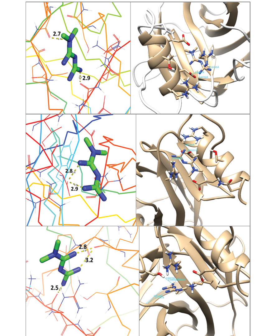
Figure 1. Three-dimensional visualization of the top binding poses from molecular docking analysis of metformin with the p53 protein (PDB ID: 1KZY). Left panels: UCSF Chimera representations showing metformin (green sticks) within the predicted binding pocket of p53, with hydrogen bond distances (Å) labeled and interacting residues color-coded by secondary structure. Right panels: PyMOL representations showing the same docked complexes, with p53 depicted as a beige cartoon and interacting side chains displayed as sticks. Hydrogen bonds are indicated by dashed lines, and distances are annotated. The results illustrate key interactions such as hydrogen bonding with Arg273 and Glu285 and hydrophobic contacts with Asp281, Val272, and Phe270, consistent with the predicted high-affinity binding pose from AutoDock Vina.
|
Aaliazar F, et al |
Investigating the Effect of Metformin on p53 Protein Using a Molecular Docking Approach |
|
6 |
GMJ.2026;15:e3872 www.salviapub.com |
|
Investigating the Effect of Metformin on p53 Protein Using a Molecular Docking Approach |
Aaliazar F, et al |
|
GMJ.2026;15:e3872 www.salviapub.com |
7 |
|
References |
|
Aaliazar F, et al |
Investigating the Effect of Metformin on p53 Protein Using a Molecular Docking Approach |
|
8 |
GMJ.2026;15:e3872 www.salviapub.com |