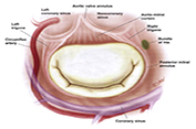
Morphometric Study of Mitral Valve Annulus in Iran
DOI:
https://doi.org/10.31661/gmj.v7i.1078Keywords:
Mitral Valve, Cadaver, Cardiac MorphologyAbstract
Background: This study aimed to determine the normal dimensions of the mitral annulus (MA) in Iranian population. Materials and Methods: This cross-sectional study was conducted using 88 fresh hearts of male and female cadavers for six months in Mashhad, Iran. Normal data were determined by measuring the exact dimensions of the MA in fresh hearts of patients who had died of non-cardiac causes and considering some parameters such as age, gender, stature, and weight. Images of the valves and leaflets were prepared by marking the anterior (A2, midpoint of anterior) and posterior areas of P1, P2, and P3 using a needle. To analyze the data, SPSS version 16 was used. Results: The means of anatomic area, anatomic perimeter, inter-commissural distance, A2-P1, A2-P2, A2-P3, Base-P1, Base-P2, Base-P3, and Base-A were 14±1.28, 8.3±1, 2.7±0.42, 2.27±0.37, 2.3±0.43, 2.06±0.35, 1.66±0.43, 1.2±0.97, 1.5±0.66, and 3.2±0.52, respectively. Comparison of the age groups regarding valve leaflets showed that Strut-P1 and Base-P2 were significantly different. Comparison of the valve leaflets and sub-valve indicators between the two genders reflected no significant differences. Age groups differed significantly in terms of Strut-P1 and Base-P2 (P=0.004 and P=00.1, respectively). Conclusions: A2-P3, A2-P1, anatomic perimeter, and anatomic area were found to be related to gender. A2-P1 and A2-P2 and some leaflet indicators such as Strut-P1 and Base-P2 were associated with age, whereas Base-P2 was affected by body mass index. [GMJ.2018;7:e1078]
References
Dwivedi G, Mahadevan G, Jimenez D, Frenneaux M, Steeds RP. Reference values for mitral and tricuspid annular dimensions using two-dimensional echocardiography. Echo Res Pract. 2014;1(2):43-50. Bahramnezhad F, Noughabi AA, Afshar PF, Marandi S. Exercise and quality of life in patients with chronic heart failure. Galen Medical Journal. 2013;2(2):49-53. Khansha R, Miladpour B, Mostafavi-Pour Z, Zal F. Lipid peroxidation product and glutathione levels in patients with coronary heart disease before and after surgery. Galen Medical Journal. 2015;4(2):78-82. Fukuda S, Watanabe H, Daimon M, Abe Y, Hirashiki A, Hirata K, et al. Normal values of real-time 3-dimensional echocardiographic parameters in a healthy Japanese population: the JAMP-3D Study. Circ J. 2012; 76(5):1177–81. Sadeghpour A, Shahrabi MR, Bakhshandeh H, Naderi N. Normal Echocardiographic Values of 368 Iranian Healthy Subjects. Arch Cardiovasc Image. 2013; 1(2):72-9. Kopuz C, Erk K, Baris YS, Onderoglu S, Sinav A. Morphometry of the fibrous ring of the mitral valve. Ann Anat. 1995; 177(2):151-4. Eto M, Morita SH, Nakashima Y, Nishimura Y, Tominaga R. Morphometric study of the human mitral annulus Guide for mitral valve surgery. Asian Cardiovasc Thorac Ann. 2013; 22(7): 787-93. Tamborini G, Fusini L, Muratori M, Gripari P, Ali SG, Fiorentini C, et al. Right heart chamber geometry and tricuspid annulus morphology in patients undergoing mitral valve repair with and without tricuspid valve annuloplasty. Int J Cardiovasc Imaging. 2016;32(6):885-94. Keller F, Werner R, Wähner J, Köhler T, Wolff W, Leutert G. Histological biomorphosis of human heart valve. II. Z Gerontol Geriatr. 1999 ; 32(2):104-11. [in German] Sonne C, Sugeng L, Watanabe N, Weinert L, Saito K, Tsukiji M, et al. Age and body surface area dependency of mitral valve and papillary apparatus parameters:assessment by real-time three-dimensional echocardiography. Eur J Echocardiogr. 2009; 10(2):287-94. Mohammadi S, Hedjazi A, Sajjadian M, Ghoroubi N, Mohammadi M, Erfani S. Study of the normal heart size in Northwest part of Iranian population: a cadaveric study. J Cardiovasc Thorac Res. 2016; 8(3):119-25.


