The Role of the Ultrasonographic Knee Assessment in the Diagnosis of Patellofemoral Pain Syndrome
DOI:
https://doi.org/10.31661/gmj.v11i.2420Keywords:
Patellofemoral Pain Syndrome, Sulcus, Q Angle, UltrasonographyAbstract
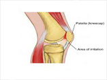
Background: Patellofemoral Pain Syndrome (PFPS) is a common anterior knee compartment pain etiology. It has been aimed to assess the ultrasonographic findings of the patellofemoral joint in patients with PFPS versus healthy individuals. Materials and Methods: The current case-control investigation was performed on 30 cases suffering from patellofemoral joint pain and 30 healthy individuals in Isfahan during 2020-21. All cases underwent ultrasonography to assess cartilage thickness, sulcus angle, and sulcus depth. We also measured the Q angle with a manual goniometer. Results: In healthy individuals, Q angle scores were statistically lower than in the cases group (P=0.002). The sulcus angle was remarkably higher among the patients compared to the controls. The cartilage thickness (P=0.88) and sulcus depth (P=0.543) scores had no statistical difference between the PFPS and healthy subjects (P<0.05). Conclusions: Patients with PFPS had significantly higher Q angle and lower sulcus angle than the healthy controls.
References
Gaitonde DY, Ericksen A, Robbins RC. Patellofemoral Pain Syndrome. Am Fam Physician. 2019;99(2):88-94. Alba-MartÃn P, Gallego-Izquierdo T, Plaza-Manzano G, Romero-Franco N, Núñez-Nagy S, Pecos-MartÃn D. Effectiveness of therapeutic physical exercise in the treatment of patellofemoral pain syndrome: a systematic review. J Phys Ther Sci. 2015;27(7):2387-90. https://doi.org/10.1589/jpts.27.2387PMid:26311988 PMCid:PMC4540887 Gulati A, McElrath C, Wadhwa V, Shah JP, Chhabra A. Current clinical, radiological and treatment perspectives of patellofemoral pain syndrome. Br J Radiol. 2018;91(1086):20170456. https://doi.org/10.1259/bjr.20170456PMid:29303366 PMCid:PMC6223269 Collins NJ, Barton CJ, Van Middelkoop M, Callaghan MJ, Rathleff MS, Vicenzino BT, et al. 2018 Consensus statement on exercise therapy and physical interventions (orthoses, taping and manual therapy) to treat patellofemoral pain: recommendations from the 5th International Patellofemoral Pain Research Retreat, Gold Coast, Australia, 2017. Br J Sports Med. 2018;52(18):1170-8. https://doi.org/10.1136/bjsports-2018-099397PMid:29925502 Rothermich MA, Glaviano NR, Li J, Hart JM. Patellofemoral pain: epidemiology, pathophysiology, and treatment options. Clin Sports Med. 2015;34(2):313-27. https://doi.org/10.1016/j.csm.2014.12.011PMid:25818716 Smith BE, Selfe J, Thacker D, Hendrick P, Bateman M, Moffatt F, et al. Incidence and prevalence of patellofemoral pain: A systematic review and meta-analysis. PLoS One. 2018;13(1):e0190892. https://doi.org/10.1371/journal.pone.0190892PMid:29324820 PMCid:PMC5764329 Glaviano NR, Kew M, Hart JM, Saliba S. Demographic and epidemiological trends in patellofemoral pain. Int J Sports Phys Ther. 2015;10(3):281-90. Tramontano M, Pagnotta S, Lunghi C, Manzo C, Manzo F, Consolo S, Manzo V. Assessment and Management of Somatic Dysfunctions in Patients With Patellofemoral Pain Syndrome. J Am Osteopath Assoc. 2020;120(3):165-73. https://doi.org/10.7556/jaoa.2020.029PMid:32091560 Cui LH. Research progress on the etiology and treatment of patellofemoral pain syndrome. Zhongguo Gu Shang. 2017;30(7):680-4. Van Der Heijden RA, Lankhorst NE, Van Linschoten R, Bierma-Zeinstra SM, Van Middelkoop M. Exercise for treating patellofemoral pain syndrome. Cochrane Database Syst Rev. 2015;1:CD010387. https://doi.org/10.1002/14651858.CD010387.pub2PMCid:PMC6457759 Ghany JF, Kamel S, Zoga A, Farrell T, Morrison W, Belair J, Desai V. Extensor mechanism tendinopathy in patients with lateral patellar maltracking. Skeletal Radiol. 2021;50(11):2205-12. https://doi.org/10.1007/s00256-021-03787-8PMid:33876276 Crossley KM, Callaghan MJ, Van Linschoten R. Patellofemoral pain. BMJ. 2015;351:h3939. https://doi.org/10.1136/bmj.h3939PMid:26537829 Drew BT, Redmond AC, Smith TO, Penny F, Conaghan PG. Which patellofemoral joint imaging features are associated with patellofemoral pain? Systematic review and meta-analysis. Osteoarthritis Cartilage. 2016;24(2):224-36. https://doi.org/10.1016/j.joca.2015.09.004PMid:26471209 Van Der Heijden RA, De Kanter JL, Bierma-Zeinstra SM, Verhaar JA, Van Veldhoven PL, Krestin GP, et al. Structural Abnormalities on Magnetic Resonance Imaging in Patients With Patellofemoral Pain: A Cross-sectional Case-Control Study. Am J Sports Med. 2016;44(9):2339-46. https://doi.org/10.1177/0363546516646107PMid:27206691 Van Middelkoop M, Macri EM, Eijkenboom JF, Van Der Heijden RA, Crossley KM, Bierma-Zeinstra SMA, et al. Are Patellofemoral Joint Alignment and Shape Associated With Structural Magnetic Resonance Imaging Abnormalities and Symptoms Among People With Patellofemoral Pain? Am J Sports Med. 2018;46(13):3217-26. https://doi.org/10.1177/0363546518801314PMid:30321064 PMCid:PMC6236631 Drew BT, Conaghan PG, Smith TO, Selfe J, Hensor EMA, Dube B, et al. Toward the Development of Data-Driven Diagnostic Subgroups for People With Patellofemoral Pain Using Modifiable Clinical, Biomechanical, and Imaging Features. J Orthop Sports Phys Ther. 2019;49(7):536-47. https://doi.org/10.2519/jospt.2019.8607PMid:31213159 Fanlo-Mazas P, Bueno-Gracia E, De Escudero-Zapico AR, Tricás-Moreno JM, Lucha-López MO. The Effect of Diacutaneous Fibrolysis on Patellar Position Measured Using Ultrasound Scanning in Patients With Patellofemoral Pain Syndrome. J Sport Rehabil. 2019;28(6):564-9. https://doi.org/10.1123/jsr.2017-0272PMid:29651911 Romero-Morales C, Bravo-Aguilar M, Ruiz-Ruiz B, Almazán-Polo J, López-López D, Blanco-Morales M, et al. Current advances and research in ultrasound imaging to the assessment and management of musculoskeletal disorders. Dis Mon. 2021;67(3):101050. https://doi.org/10.1016/j.disamonth.2020.101050PMid:32711897 Jeon H, Donovan L, Thomas AC. Exercise-Induced Changes in Femoral Cartilage Thickness in Patients with Patellofemoral Pain. J Athl Train (in press). 2022. https://doi.org/10.4085/1062-6050-0602.21PMid:35476136 Brushøj C, Hölmich P, Nielsen MB, Albrecht-Beste E. Acute patellofemoral pain: aggravating activities, clinical examination, MRI and ultrasound findings. Br J Sports Med. 2008;42(1):64-7. https://doi.org/10.1136/bjsm.2006.034215PMid:17562742 Lin YF, Lin JJ, Cheng CK, Lin DH, Jan MH. Association between sonographic morphology of vastus medialis obliquus and patellar alignment in patients with patellofemoral pain syndrome. J Orthop Sports Phys Ther. 2008;38(4):196-202. https://doi.org/10.2519/jospt.2008.2568PMid:18434663 Payne K, Payne J, Larkin TA. Patellofemoral Pain Syndrome and Pain Severity Is Associated With Asymmetry of Gluteus Medius Muscle Activation Measured Via Ultrasound. Am J Phys Med Rehabil. 2020;99(7):595-601. https://doi.org/10.1097/PHM.0000000000001367PMid:31860586 Schoots EJ, Tak IJ, Veenstra BJ, Krebbers YM, Bax JG. Ultrasound characteristics of the lateral retinaculum in 10 patients with patellofemoral pain syndrome compared to healthy controls. J Bodyw Mov Ther. 2013;17(4):523-9. https://doi.org/10.1016/j.jbmt.2013.03.005PMid:24139014 Fischhoff C. Patellofemoral pain syndrome: ultrasound measurements for diagnosis. Int Musculoskelet Med. 2015;37(2):54-8. https://doi.org/10.1179/1753614615Z.000000000100