
Micro-CT Evaluation of Marginal and Internal Fit of Lithium Disilicate Crowns – Influence of Wax-Up and Manufacturing Technique
DOI:
https://doi.org/10.31661/gmj.v13iSP1.3562Keywords:
Lithium Disilicate; Crown; Milling; 3DprintAbstract
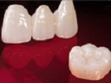
Background: Lithium disilicate crowns are widely used in dentistry, with various fabrication methods available. However, there is a research gap in comparing the marginal and internal fit of these crowns produced through different manufacturing techniques. This study aims to evaluate the impact of various manufacturing methods on the marginal and internal fit of lithium disilicate crowns. Materials and Methods: The left maxillary canine's acrylic tooth was prepared using a high-speed handpiece. Subsequently, the prepared tooth was scanned with a laboratory scanner, and 40 dies were milled with resin. These dies were then divided into four groups (n=10), with lithium disilicate crowns fabricated using different methods for each group: group 1) conventional wax-up method and heat press, group 2) 3D print wax-up method and heat press, group 3) wax-up milling method and heat press, and group 4) CAD/CAM method. The marginal and internal fit of the crowns were assessed using micro-CT by measuring Absolute Marginal Discrepancy (AMD), Marginal Gap (MG), Axial Gap (AG), and Occlusal Gap (OG) at various points. Group comparisons were conducted using one-way ANOVA, while Spearman rank correlation coefficient was used to evaluate variable correlations (α=0.05). Results: ANOVA analysis indicated significant differences among all groups for most examined points except for lingual AMD. In inter-group comparisons, the CAD/CAM method demonstrated superior results in MG buccal, MG lingual, AMD buccal, AG2, AG3, OG1, OG3, and OG4 measurements. The 3D printing method outperformed in AG1 and OG2 comparisons while the milling method excelled in AG4 comparison. Although no significant difference was observed in lingual AMD comparison among groups, the CAD/CAM approach exhibited better average agreement. Overall, the conventional group displayed the weakest performance in terms of adaptation. Conclusions: The study findings suggest that all-digital and semi-digital methods for fabricating lithium disilicate crowns offer better adaptation compared to conventional techniques.
References
Sailer I, Makarov NA, Thoma DS, Zwahlen M, Pjetursson BE. All-ceramic or metal-ceramic tooth-supported fixed dental prostheses (FDPs)? A systematic review of the survival and complication rates. Part I: Single crowns (SCs). Dent Mater 2015;31:603-23.
https://doi.org/10.1016/j.dental.2015.02.011
PMid:25842099
Gardner FM. Margins of complete crowns--literature review. J Prosthet Dent. 1982;48:396-400.
https://doi.org/10.1016/0022-3913(82)90072-5
PMid:6752383
Pettenò D, Schierano G, Bassi F, Bresciano ME, Carossa S. Comparison of marginal fit of 3 different metal-ceramic systems: an in vitro study. Int J Prosthodont. 2000;13:405-8.
Barão VA, Gennari-Filho H, Goiato MC, dos Santos DM, Pesqueira AA. Factors to achieve aesthetics in all-ceramic restorations. J Craniofac Surg. 2010;21:2007-12.
https://doi.org/10.1097/SCS.0b013e3181f535d4
PMid:21119487
Rauch A, Reich S, Dalchau L, Schierz O. Clinical survival of chair-side generated monolithic lithium disilicate crowns:10-year results. Clin Oral Investig. 2018;22:1763-9.
https://doi.org/10.1007/s00784-017-2271-3
PMid:29103104
Warreth A, Elkareimi Y. All-ceramic restorations: A review of the literature. Saudi Dent J. 2020;32:365-72.
https://doi.org/10.1016/j.sdentj.2020.05.004
PMid:34588757 PMCid:PMC8461086
Teichmann M, Göckler F, Weber V, Yildirim M, Wolfart S, Edelhoff D. Ten-year survival and complication rates of lithium-disilicate (Empress 2) tooth-supported crowns, implant-supported crowns, and fixed dental prostheses. J Dent. 2017;56:65-77.
https://doi.org/10.1016/j.jdent.2016.10.017
PMid:27984088
Teichmann M, Göckler F, Rückbeil M, Weber V, Edelhoff D, Wolfart S. Periodontal outcome and additional clinical quality criteria of lithium-disilicate restorations (Empress 2) after 14 years. Clin Oral Investig. 2019;23:2153-64.
https://doi.org/10.1007/s00784-018-2649-x
PMid:30276515
Willard A, Gabriel Chu T-M. The science and application of IPS e.Max dental ceramic. The Kaohsiung Journal of Medical Sciences. 2018;34:238-42.
https://doi.org/10.1016/j.kjms.2018.01.012
PMid:29655413
Potiket N, Chiche G, Finger IM. In vitro fracture strength of teeth restored with different all-ceramic crown systems. J Prosthet Dent. 2004;92:491-5.
https://doi.org/10.1016/j.prosdent.2004.09.001
PMid:15523339
Odén A, Andersson M, Krystek-Ondracek I, Magnusson D. Five-year clinical evaluation of Procera AllCeram crowns. J Prosthet Dent. 1998;80:450-6.
https://doi.org/10.1016/S0022-3913(98)70010-1
PMid:9791792
Rosenstiel SF MB, Land MF, Msd D, Walter R. Contemporary Fixed Prosthodontics-e-book Elsevier Health Sciences.6th ed. Philadelphia: Elsivier; 2022.
Selukar M, Godbole Dubey S, Sathe Kambla S. Comparative evaluation of marginal fit, internal fit and cement space of single unit lithium disilicate crown with shoulder finish line by two different methods of fabrication. F1000Research. 2023;12:1510.
https://doi.org/10.12688/f1000research.135050.1
Höland W, Schweiger M, Frank M, Rheinberger V. A comparison of the microstructure and properties of the IPS Empress 2 and the IPS Empress glass-ceramics. J Biomed Mater Res. 2000;53:297-303.
https://doi.org/10.1002/1097-4636(2000)53:4<297::AID-JBM3>3.0.CO;2-G
PMid:10898870
Jalalian E, Moghadam L. Compare the fracture resistance of 2 All ceramic systems, IPS e.max, IPS Empress. J Dent (Shiraz University) . 2008;9:51-7.
Guess PC, Schultheis S, Bonfante EA, Coelho PG, Ferencz JL, Silva NR. All-ceramic systems: laboratory and clinical performance. Dent Clin North Am. 2011;55:333-52.
https://doi.org/10.1016/j.cden.2011.01.005
PMid:21473997
Christensen GJ. Marginal fit of gold inlay castings. J Prosthet Dent. 1966;16:297-305.
https://doi.org/10.1016/0022-3913(66)90082-5
PMid:5217112
McLean JW, von Fraunhofer JA. The estimation of cement film thickness by an in vivo technique. Br Dent J. 1971;131:107-11.
https://doi.org/10.1038/sj.bdj.4802708
PMid:5283545
Vargas-Corral FG, Vargas-Corral AE, Valverde MAR, Bravo M, Leal JIR. Clinical comparison of marginal fit of ceramic inlays between digital and conventional impressions. J Adv Prosthodont. 2024;16:57-65.
https://doi.org/10.4047/jap.2024.16.1.57
PMid:38455677 PMCid:PMC10917630
Tsirogiannis P, Reissmann DR, Heydecke G. Evaluation of the marginal fit of single-unit, complete-coverage ceramic restorations fabricated after digital and conventional impressions: A systematic review and meta-analysis. J Prosthet Dent. 2016;116:328-35.e2.
https://doi.org/10.1016/j.prosdent.2016.01.028
PMid:27061627
Goujat A, Abouelleil H, Colon P, Jeannin C, Pradelle N, Seux D, et al. Marginal and internal fit of CAD-CAM inlay/onlay restorations: A systematic review of in vitro studies. J Prosthet Dent. 2019;121:590-7.e3.
https://doi.org/10.1016/j.prosdent.2018.06.006
PMid:30509548
Bindl A, Mörmann WH. Marginal and internal fit of all-ceramic CAD/CAM crown-copings on chamfer preparations. J Oral Rehabil. 2005;32(6):441-7.
https://doi.org/10.1111/j.1365-2842.2005.01446.x
PMid:15899023
Ferrairo BM, Piras FF, Lima FF, Honório HM, Duarte MAH, Borges AFS, et al. Comparison of marginal adaptation and internal fit of monolithic lithium disilicate crowns produced by 4 different CAD/CAM systems. Clin Oral Investig. 2021;25:2029-36.
https://doi.org/10.1007/s00784-020-03511-1
PMid:32783095
Conrad HJ, Seong WJ, Pesun IJ. Current ceramic materials and systems with clinical recommendations: a systematic review. J Prosthet Dent. 2007;98:389-404.
https://doi.org/10.1016/S0022-3913(07)60124-3
PMid:18021828
Zarone F, Di Mauro MI, Ausiello P, Ruggiero G, Sorrentino R. Current status on lithium disilicate and zirconia: a narrative review. BMC oral health. 2019;19:1-14.
https://doi.org/10.1186/s12903-019-0838-x
PMid:31272441 PMCid:PMC6610968
Fabian Fonzar R, Carrabba M, Sedda M, Ferrari M, Goracci C, Vichi A. Flexural resistance of heat-pressed and CAD-CAM lithium disilicate with different translucencies. Dental Materials. 2017;33:63-70.
https://doi.org/10.1016/j.dental.2016.10.005
PMid:27855994
Alghazzawi TF, Liu PR, Essig ME. The effect of different fabrication steps on the marginal adaptation of two types of glass-infiltrated ceramic crown copings fabricated by CAD/CAM technology. J Prosthodont. 2012;21:167-72.
https://doi.org/10.1111/j.1532-849X.2011.00803.x
PMid:22372838
Lin MT, Sy-Muñoz J, Muñoz CA, Goodacre CJ, Naylor WP. The effect of tooth preparation form on the fit of Procera copings. Int J Prosthodont. 1998;11:580-90.
Reich S, Wichmann M, Nkenke E, Proeschel P. Clinical fit of all-ceramic three-unit fixed partial dentures, generated with three different CAD/CAM systems. Eur J Oral Sci. 2005;113:174-9.
https://doi.org/10.1111/j.1600-0722.2004.00197.x
PMid:15819826
Silva NR, Witek L, Coelho PG, Thompson VP, Rekow ED, Smay J. Additive CAD/CAM process for dental prostheses. J Prosthodont. 2011;20:93-6.
https://doi.org/10.1111/j.1532-849X.2010.00623.x
PMid:20561158
Abduo J, Lyons K, Bennamoun M. Trends in computer-aided manufacturing in prosthodontics: a review of the available streams. Int J Dent. 2014;2014:783948.
https://doi.org/10.1155/2014/783948
PMid:24817888 PMCid:PMC4000974
Arezoobakhsh A, Shayegh SS, Jamali Ghomi A, Hakimaneh SMR. Comparison of marginal and internal fit of 3-unit zirconia frameworks fabricated with CAD-CAM technology using direct and indirect digital scans. J Prosthet Dent. 2020;123:105-12.
https://doi.org/10.1016/j.prosdent.2018.10.023
PMid:30982618
Mously HA, Finkelman M, Zandparsa R, Hirayama H. Marginal and internal adaptation of ceramic crown restorations fabricated with CAD/CAM technology and the heat-press technique. J Prosthet Dent. 2014;112:249-56.
https://doi.org/10.1016/j.prosdent.2014.03.017
PMid:24795263
Shamseddine L, Mortada R, Rifai K, Chidiac JJ. Marginal and internal fit of pressed ceramic crowns made from conventional and computer-aided design and computer-aided manufacturing wax patterns: An in vitro comparison. J Prosthet Dent. 2016;116:242-8.
https://doi.org/10.1016/j.prosdent.2015.12.005
PMid:26948080
Fathi HM, Al-Masoody AH, El-Ghezawi N, Johnson A. The Accuracy of Fit of Crowns Made From Wax Patterns Produced Conventionally (Hand Formed) and Via CAD/CAM Technology. Eur J Prosthodont Restor Dent. 2016;24:10-7.
Homsy FR, Özcan M, Khoury M, Majzoub ZAK. Marginal and internal fit of pressed lithium disilicate inlays fabricated with milling, 3D printing, and conventional technologies. J Prosthet Dent. 2018;119:783-90.
https://doi.org/10.1016/j.prosdent.2017.07.025
PMid:28969918
Mostafa NZ, Ruse ND, Ford NL, Carvalho RM, Wyatt CCL. Marginal Fit of Lithium Disilicate Crowns Fabricated Using Conventional and Digital Methodology: A Three-Dimensional Analysis. J Prosthodont. 2018;27:145-52.
https://doi.org/10.1111/jopr.12656
PMid:28833920
Al Hamad KQ, Al Quran FA, AlJalam SA, Baba NZ. Comparison of the Accuracy of Fit of Metal, Zirconia, and Lithium Disilicate Crowns Made from Different Manufacturing Techniques. J Prosthodont. 2019;28:497-503.
https://doi.org/10.1111/jopr.13029
PMid:30719780
Dolev E, Bitterman Y, Meirowitz A. Comparison of marginal fit between CAD-CAM and hot-press lithium disilicate crowns. J Prosthet Dent. 2019;121:124-8.
https://doi.org/10.1016/j.prosdent.2018.03.035
PMid:29961628
Lim YA, Kim JM, Choi Y, Park S. Evaluation of Fitness and Accuracy of Milled and Three-Dimensionally Printed Inlays. Eur J Dent. 2023;17:1029-36.
https://doi.org/10.1055/s-0042-1758796
PMid:36599450 PMCid:PMC10756847
Published
How to Cite
Issue
Section
License
Copyright (c) 2024 Galen Medical Journal

This work is licensed under a Creative Commons Attribution 4.0 International License.


