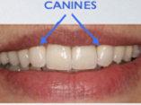
Canine Tooth Position and its Relationship with the Perpendicular Line to the Nasal Ala in an Iranian Population
DOI:
https://doi.org/10.31661/gmj.v13iSP1.3568Keywords:
Anatomic Landmarks; Cuspid; Esthetics, Dental; Nasal CartilagesAbstract
Background: Position of anterior teeth is an important step in smile design and fabrication of full dentures. This study aimed to assess the canine tooth position and its relationship with the line drawn perpendicular to the nasal ala in an Iranian population. Materials and Method: This analytical epidemiological field study was conducted on 100 males and 100 females presenting to the dental clinic of Shahed Dental School in June 2023. Digital photographs were obtained from the patients under standardized conditions. Reference lines were drawn perpendicular to the nasal alae and continued down to the lower lip (AI lines). The distance from the canine cusp tip (AICT) and distal contact point of the canine tooth (AICD) to the AI line was measured bilaterally using Digimizer software version 6. Data were analyzed by t-test (alpha=0.05). Results: The AICT and AICD were significantly larger than zero in both males and females (P<0.05). The mean AICT and AICD in males were larger than those in females by 2-2.5 mm. No significant difference was found in the right and left canine position, neither in males nor in females (P>0.05). Conclusion: According to the results, the AI line and gender both affected the canine position in the study population, and therefore, should be taken into account in designing an esthetically pleasant denture. Canine position properly determined for one side may be used for the other side as well since no significant difference was found in the right and left canine position.
References
Proffit WR, Fields H, Msd DM, Larson B, Sarver DM. Contemporary Orthodontics, 6e. South Asia Edition-E-Book: Elsevier India; 2019.
Dindaroğlu F, Ertan Erdinç AM, Doğan S. Perception of Smile Esthetics by Orthodontists and Laypersons: Full Face and A Localized View of The Social and Spontaneous Smiles. Turk J Orthod. 2016 Sep;29(3):59-68.
https://doi.org/10.5152/TurkJOrthod.2016.0013
PMid:30112476 PMCid:PMC6007622
Öz AA, Akdeniz BS, Canlı E, Çelik S. Smile Attractiveness: Differences among the Perceptions of Dental Professionals and Laypersons. Turk J Orthod. 2017 Jun;30(2):50-55.
https://doi.org/10.5152/TurkJOrthod.2017.17021
PMid:30112492 PMCid:PMC6007756
Batwa W. The Influence of the Smile on the Perceived Facial Type Esthetics. Biomed Res Int. 2018 Jul 9;2018:3562916.
https://doi.org/10.1155/2018/3562916
PMid:30112381 PMCid:PMC6077656
Jacobson A, White L. Radiographic cephalometry: from basics to 3-D imaging. American Journal of Orthodontics and Dentofacial Orthopedics. 2007 Apr 1;131(4):S133.
https://doi.org/10.1016/j.ajodo.2007.02.038
Heravi F, Ahrari F, Rashed R, Heravi P, Ghaffari N, Habibirad A. Evaluation of factors affecting dental esthetics in patients seeking orthodontic treatment. Int J Orthod Rehabil. 2016 Jan 12;7(3):79-84.
https://doi.org/10.4103/2349-5243.192526
Jacobson A. Color atlas of dental medicine Orthodontic-Diagnosis: Thomas Rakosi, Irmtrud Jonas and Thomas M. Am J Orthod Dentofacial Orthop. 1994 Jun 1;105(6):613.
https://doi.org/10.1016/S0889-5406(05)80780-2
Omrani A, Barekatain M, Hasanli E, Abdolmaleki M. Comparison of anthropometry and digital photography techniques in facial proportion analysis. J Isfahan Dent Sch. 2012 Dec 1;8(5):453-62.
Pandey AK. Cephalo-facial variation among Onges. Anthropol. 2006 Oct 1;8(4):245-9.
https://doi.org/10.1080/09720073.2006.11890971
Hasanreisoglu U, Berksun S, Aras K, Arslan I. An analysis of maxillary anterior teeth: facial and dental proportions. J Prosthet Dent. 2005 Dec;94(6):530-8.
https://doi.org/10.1016/j.prosdent.2005.10.007
PMid:16316799
Alavi S, Safari A. An investigation on facial and cranial anthropometric parameters among Isfahan Young adults. J Dent Med. 2003 Apr 10;16(1):19-28.
Shetti VR, Pai SR, Sneha GK, Gupta C, Chethan P. Estudio del Índice prosopo (facial) de estudiantes de la India y Malasia. Int J Morphol. 2011 Sep;29(3):1018-21.
https://doi.org/10.4067/S0717-95022011000300060
Khane Masjedi M, Basir L, Nourollah M. The Relationship between Facial Index and Smile Features in People Aged 20-35 in Ahvaz. Jundishapur J Med Sci. 2022; 20(Special Issue):652-63.
Nasiri P, Estebsari A, Hosseini Shirazi SM, Mokhtarpour H, Mousavi SJ, Ebrahimi Saravi M. Relationship between anterior teeth sizes and facial dimensions in dentate patients. J Maz Univ Med Sci. 2021 Feb 10;30(194):100-7.
Tarfa SJ, Salih NF. Relationship among inter canine width, intermolar width, and arch length in upper and lower arch for dentistry students of Thi-Qar University. J Res Med Dent Sci. 2021;9:126-30.
Pisulkar S, Nimonkar S, Bansod A, Belkhode V, Godbole S. Quantifying the Selection of Maxillary Anterior Teeth Using Extraoral Anatomical Landmarks. Cureus. 2022 Jul 28;14(7):e27410.
https://doi.org/10.7759/cureus.27410
Srimaneekarn N, Arayapisit T, Pooktuantong O, Cheng HR, Soonsawad P. Determining canine position by multiple facial landmarks to achieve natural esthetics in complete denture treatment. J Prosthet Dent. 2022 Jun;127(6):860-5.
https://doi.org/10.1016/j.prosdent.2020.11.022
PMid:33468316 PMCid:PMC8282807
Bonakdarchian M, Ghorbanipour R. The relationship between the nose blades and the size of artificial teeth. J Isfahan Dent Sch. 2010;6(3):207-13.

Published
How to Cite
Issue
Section
License
Copyright (c) 2024 Galen Medical Journal

This work is licensed under a Creative Commons Attribution 4.0 International License.


