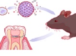
Biocompatibility of A Nano-curcumin Pulpal Paste in Rats
DOI:
https://doi.org/10.31661/gmj.v13iSP1.3579Keywords:
Materials Testing; Curcumin; Metapex; Rats; Tooth; DeciduousAbstract
Background: This study aimed to assess the biocompatibility of different concentrations of a nano-curcumin pulpal paste in rats. Materials and Methods: Polyethylene tubes containing zinc oxide eugenol (ZOE), Metapex, and 2, 4, 6, and 8 ppm nano-curcumin pulpal paste, and an empty tube as the negative control were implanted in the back of 30 Wistar rats (7 tubes per each rat). The rats were sacrificed after 15, 30, and 60 days (10 rats at each time point). The tissue around the tubes underwent histopathological analysis. After hematoxylin and eosin staining, the specimens were evaluated for presence/absence of necrosis and calcification, number of inflammatory cells, and thickness of soft tissue capsule. Data were analyzed by the Chi-square, Mann-Whitney, and Kruskal-Wallis tests (α=0.05). Results: Necrosis was not seen in any group at any time point. No significant difference existed among the experimental groups regarding calcification at different time points (P>0.05). The fibrotic capsule was thin in all experimental groups at all time points. Rate of inflammation decreased in all experimental groups from day 15 to day 60. Among different concentrations, only 2 ppm concentration of nano-curcumin paste had no significant difference with Metapex and ZOE regarding inflammation at different time points. Conclusion: All tested concentrations of nano-curcumin pulpal paste were biocompatible, compared with the positive controls (ZOE and Metapex); but 2 ppm concentration was the most biocompatible. Within the limitations of this in vitro study, 2 ppm concentration of nano-curcumin may be suggested for further experiments.
References
Dummett CO, Kopel HM. Pediatric Endodontics. In Ingle and Bakland. 5th ed. Endodontics: B.C. Decker Elsevier; 2002. (pp. 861-902).
Silva LA, Leonardo MR, Oliveira DS, Silva RA, Queiroz AM, Hernandez PG, et al. Histopathological evaluation of root canal filling materials for primDummett CO, Kopel HM. Pediatric Endodontics In Ingle and Bakland 5th ed Endodontics: BC Decker. Elsevier. 2002;:861-902.
Silva LA, Leonardo MR, Oliveira DS, Silva RA, Queiroz AM, Hernandez PG, et al. Histopathological evaluation of root canal filling materials for primary teeth. Brazilian dental journal. 2010;21(1):38-45.
https://doi.org/10.1590/S0103-64402010000100006
PMid:20464319
Fuks A, Peretz B. Pediatric endodontics: current concepts in pulp therapy for primary and young permanent teeth 2nd ed. Springer International Publishing Switzerland. 2016; : 83.
Segato RA, Pucinelli CM, Ferreira DC, Daldegan Ade R, Silva RS, Nelson-Filho P, et al. Physicochemical Properties of Root Canal Filling Materials for Primary Teeth. Brazilian dental journal. 2016;27(2):196-201.
https://doi.org/10.1590/0103-6440201600206
PMid:27058384
Barja-Fidalgo F, Moutinho-Ribeiro M, Oliveira MA, de Oliveira BH. A systematic review of root canal filling materials for deciduous teeth: is there an alternative for zinc oxide-eugenol?. ISRN dentistry. 2011;2011:367318.
https://doi.org/10.5402/2011/367318
PMid:21991471 PMCid:PMC3169841
Praveen P, Anantharaj A, Venkataraghavan K, Rani P, Sudhir R, Jaya AR. A review of obturating materials for primary teeth. SRM Journal of Research in Dental Sciences. 2011;2(1):42-44.
https://doi.org/10.4103/0976-433X.121727
Palma M, Pineiro Z, Barroso CG. Stability of phenolic compounds during extraction with superheated solvents. Journal of chromatography A. 2001;921(2):169-74.
https://doi.org/10.1016/S0021-9673(01)00882-2
PMid:11471800
Joe B, Vijaykumar M, Lokesh BR. Biological properties of curcumin-cellular and molecular mechanisms of action. Critical reviews in food science and nutrition. 2004;44(2):97-111.
https://doi.org/10.1080/10408690490424702
PMid:15116757
Tonnesen HH, Karlsen J. Studies on curcumin and curcuminoids VI Kinetics of curcumin degradation in aqueous solution. Zeitschrift fur Lebensmittel-Untersuchung und -Forschung. 1985;180(5):402-4.
https://doi.org/10.1007/BF01027775
PMid:4013525
Lopez-Lazaro M. Anticancer and carcinogenic properties of curcumin: considerations for its clinical development as a cancer chemopreventive and chemotherapeutic agent. Molecular nutrition & food research. 2008;52 Suppl 1:S103-27.
https://doi.org/10.1002/mnfr.200700238
PMid:18496811
Cleary K, McFeeters RF. Effects of oxygen and turmeric on the formation of oxidative aldehydes in fresh-pack dill pickles. Journal of agricultural and food chemistry. 2006;54(9):3421-7.
https://doi.org/10.1021/jf052868k
PMid:16637703
Gowda NK, Ledoux DR, Rottinghaus GE, Bermudez AJ, Chen YC. Efficacy of turmeric (Curcuma longa), containing a known level of curcumin, and a hydrated sodium calcium aluminosilicate to ameliorate the adverse effects of aflatoxin in broiler chicks. Poultry science. 2008;87(6):1125-30.
https://doi.org/10.3382/ps.2007-00313
PMid:18493001
Priyadarsini KI. Free radical reactions of curcumin in membrane models. Free radical biology & medicine. 1997;23(6):838-43.
https://doi.org/10.1016/S0891-5849(97)00026-9
PMid:9378362
Mehanny M, Hathout RM, Geneidi AS, Mansour S. Exploring the use of nanocarrier systems to deliver the magical molecule; Curcumin and its derivatives. Journal of controlled release: official journal of the Controlled Release Society. 2016;225:1-30.
https://doi.org/10.1016/j.jconrel.2016.01.018
PMid:26778694
Al-Rohaimi AH. Comparative anti-inflammatory potential of crystalline and amorphous nano curcumin in topical drug delivery. Journal of oleo science. 2015;64(1):27-40.
https://doi.org/10.5650/jos.ess14175
PMid:25519291
Arunraj TR, Sanoj Rejinold N, Mangalathillam S, Saroj S, Biswas R, Jayakumar R. Synthesis, characterization and biological activities of curcumin nanospheres. Journal of biomedical nanotechnology. 2014;10(2):238-50.
https://doi.org/10.1166/jbn.2014.1786
PMid:24738332
Grynkiewicz G, Slifirski P. Curcumin and curcuminoids in quest for medicinal status. Acta biochimica Polonica. 2012;59(2):201-12.
https://doi.org/10.18388/abp.2012_2139
PMid:22590694
Tsai YM, Jan WC, Chien CF, Lee WC, Lin LC, Tsai TH. Optimised nano-formulation on the bioavailability of hydrophobic polyphenol, curcumin, in freely-moving rats. Food chemistry. 2011 1;127(3):918-25.
https://doi.org/10.1016/j.foodchem.2011.01.059
PMid:25214079
Bhawana B, Basniwal RK, Buttar HS, Jain VK, Jain N. Curcumin nanoparticles: preparation, characterization, and antimicrobial study. Journal of agricultural and food chemistry. 2011;59(5):2056-61.
https://doi.org/10.1021/jf104402t
PMid:21322563
Lima CC, Conde Junior AM, Rizzo MS, Moura RD, Moura MS, Lima MD, et al. Biocompatibility of root filling pastes used in primary teeth. International endodontic journal. 2015;48(5):405-16.
https://doi.org/10.1111/iej.12328
PMid:24889680
Scarparo RK, Grecca FS, Fachin EV. Analysis of tissue reactions to methacrylate resin-based, epoxy resin-based, and zinc oxide-eugenol endodontic sealers. Journal of endodontics. 2009;35(2):229-32.
https://doi.org/10.1016/j.joen.2008.10.025
PMid:19166779
Pilownic KJ, Gomes APN, Wang ZJ, Almeida LHS, Romano AR, Shen Y, et al. Physicochemical and Biological Evaluation of Endodontic Filling Materials for Primary Teeth. Brazilian dental journal. 2017;28(5):578-86.
https://doi.org/10.1590/0103-6440201701573
PMid:29215682
Costa CA, Teixeira HM, do Nascimento AB, Hebling J. Biocompatibility of two current adhesive resins. Journal of endodontics. 2000;26(9):512-6.
https://doi.org/10.1097/00004770-200009000-00006
PMid:11199790
Queiroz AM, Assed S, Consolaro A, Nelson-Filho P, Leonardo MR, Silva RA, et al. Subcutaneous connective tissue response to primary root canal filling materials. Brazilian dental journal. 2011;22(3):203-11.
https://doi.org/10.1590/S0103-64402011000300005
PMid:21915517
Molloy D, Goldman M, White RR, Kabani S. Comparative tissue tolerance of a new endodontic sealer. Oral surgery, oral medicine, and oral pathology. 1992;73(4):490-3.
https://doi.org/10.1016/0030-4220(92)90332-K
PMid:1533448
Bodrumlu E, Muglali M, Sumer M, Guvenc T. The response of subcutaneous connective tissue to a new endodontic filling material. Journal of biomedical materials research Part B, Applied biomaterials. 2008;84(2):463-7.
https://doi.org/10.1002/jbm.b.30892
PMid:17621641
Farhad AR, Hasheminia S, Razavi S, Feizi M. Histopathologic evaluation of subcutaneous tissue response to three endodontic sealers in rats. Journal of oral science. 2011;53(1):15-21.
https://doi.org/10.2334/josnusd.53.15
PMid:21467810
Yaltirik M, Ozbas H, Bilgic B, Issever H. Reactions of connective tissue to mineral trioxide aggregate and amalgam. Journal of endodontics. 2004;30(2):95-9.
https://doi.org/10.1097/00004770-200402000-00008
PMid:14977305
Al-Ostwani AO, Al-Monaqel BM, Al-Tinawi MK. A clinical and radiographic study of four different root canal fillings in primary molars. Journal of the Indian Society of Pedodontics and Preventive Dentistry. 2016;34(1):55-9.
https://doi.org/10.4103/0970-4388.175515
PMid:26838149
Gupta S, Das G. Clinical and radiographic evaluation of zinc oxide eugenol and metapex in root canal treatment of primary teeth. Journal of the Indian Society of Pedodontics and Preventive Dentistry. 2011;29(3):222-8.
https://doi.org/10.4103/0970-4388.85829
PMid:21985878
Reddy S, Ramakrishna Y. Evaluation of antimicrobial efficacy of various root canal filling materials used in primary teeth: a microbiological study. The Journal of clinical pediatric dentistry. 2007 Spring;31(3):193-8.
https://doi.org/10.17796/jcpd.31.3.t73r4061424j2578
PMid:17550046
Onay EO, Ungor M, Ozdemir BH. In vivo evaluation of the biocompatibility of a new resin-based obturation system. Oral surgery, oral medicine, oral pathology, oral radiology, and endodontics. 2007;104(3):e60-6.
https://doi.org/10.1016/j.tripleo.2007.03.006
PMid:17618139
Olsson B, Sliwkowski A, Langeland K. Subcutaneous implantation for the biological evaluation of endodontic materials. Journal of Endodontics. 1981;7(8):355-69.
https://doi.org/10.1016/S0099-2399(81)80057-X
PMid:7021745
Mandrol PS, Bhat K, Prabhakar AR. An in vitro evaluation of cytotoxicity of curcumin against human dental pulp fibroblasts. Journal of the Indian Society of Pedodontics and Preventive Dentistry. 2016;34(3):269-72.
https://doi.org/10.4103/0970-4388.186757
PMid:27461812
Hugar SM, Kukreja P, Hugar SS, Gokhale N, Assudani H. Comparative Evaluation of Clinical and Radiographic Success of Formocresol, Propolis, Turmeric Gel, and Calcium Hydroxide on Pulpotomized Primary Molars: A Preliminary Study. International journal of clinical pediatric dentistry. 2017;10(1):18-23.
https://doi.org/10.5005/jp-journals-10005-1400
PMid:28377649 PMCid:PMC5360797
Purohit RN, Bhatt M, Purohit K, Acharya J, Kumar R, Garg R. Clinical and Radiological Evaluation of Turmeric Powder as a Pulpotomy Medicament in Primary Teeth: An in vivo Study. International journal of clinical pediatric dentistry. 2017;10(1):37-40.
https://doi.org/10.5005/jp-journals-10005-1404
PMid:28377653 PMCid:PMC5360801
Prabhakar AR, Mandroli PS, Bhat K. Pulpotomy with curcumin: Histological comparison with mineral trioxide aggregate in rats. Indian journal of dental research : official publication of Indian Society for Dental Research. 2019;30(1):31-6.
Anand P, Nair HB, Sung B, Kunnumakkara AB, Yadav VR, Tekmal RR, Aggarwal BB. Design of curcumin-loaded PLGA nanoparticles formulation with enhanced cellular uptake, and increased bioactivity in vitro and superior bioavailability in vivo. Biochemical pharmacology. 2010;79(3):330-8.
https://doi.org/10.1016/j.bcp.2009.09.003
PMid:19735646 PMCid:PMC3181156
Mohanty C, Sahoo SK. Curcumin and its topical formulations for wound healing applications. Drug discovery today. 2017;22(10):1582-92.
https://doi.org/10.1016/j.drudis.2017.07.001
PMid:28711364
Sanders JE, Rochefort JR. Fibrous encapsulation of single polymer microfibers depends on their vertical dimension in subcutaneous tissue. Journal of biomedical materials research Part A. 2003;67(4):1181-7.
https://doi.org/10.1002/jbm.a.20027
PMid:14624504
ary teeth. Brazilian dental journal. 2010;21(1):38-45.
Fuks A, Peretz B, editors. Pediatric endodontics: current concepts in pulp therapy for primary and young permanent teeth. 2nd ed. Springer International Publishing Switzerland, 2016.p 83.
Segato RA, Pucinelli CM, Ferreira DC, Daldegan Ade R, Silva RS, Nelson-Filho P, et al. Physicochemical Properties of Root Canal Filling Materials for Primary Teeth. Brazilian dental journal. 2016;27(2):196-201.
Barja-Fidalgo F, Moutinho-Ribeiro M, Oliveira MA, de Oliveira BH. A systematic review of root canal filling materials for deciduous teeth: is there an alternative for zinc oxide-eugenol?. ISRN dentistry. 2011;2011:367318.
Praveen P, Anantharaj A, Venkataraghavan K, Rani P, Sudhir R, Jaya AR. A review of obturating materials for primary teeth. SRM Journal of Research in Dental Sciences. 2011;2(1):42-44.
Palma M, Pineiro Z, Barroso CG. Stability of phenolic compounds during extraction with superheated solvents. Journal of chromatography A. 2001;921(2):169-74.
Joe B, Vijaykumar M, Lokesh BR. Biological properties of curcumin-cellular and molecular mechanisms of action. Critical reviews in food science and nutrition. 2004;44(2):97-111.
Tonnesen HH, Karlsen J. Studies on curcumin and curcuminoids. VI. Kinetics of curcumin degradation in aqueous solution. Zeitschrift fur Lebensmittel-Untersuchung und -Forschung. 1985;180(5):402-4.
Lopez-Lazaro M. Anticancer and carcinogenic properties of curcumin: considerations for its clinical development as a cancer chemopreventive and chemotherapeutic agent. Molecular nutrition & food research. 2008;52 Suppl 1:S103-27.
Cleary K, McFeeters RF. Effects of oxygen and turmeric on the formation of oxidative aldehydes in fresh-pack dill pickles. Journal of agricultural and food chemistry. 2006;54(9):3421-7.
Gowda NK, Ledoux DR, Rottinghaus GE, Bermudez AJ, Chen YC. Efficacy of turmeric (Curcuma longa), containing a known level of curcumin, and a hydrated sodium calcium aluminosilicate to ameliorate the adverse effects of aflatoxin in broiler chicks. Poultry science. 2008;87(6):1125-30.
Priyadarsini KI. Free radical reactions of curcumin in membrane models. Free radical biology & medicine. 1997;23(6):838-43.
Mehanny M, Hathout RM, Geneidi AS, Mansour S. Exploring the use of nanocarrier systems to deliver the magical molecule; Curcumin and its derivatives. Journal of controlled release: official journal of the Controlled Release Society. 2016;225:1-30.
Al-Rohaimi AH. Comparative anti-inflammatory potential of crystalline and amorphous nano curcumin in topical drug delivery. Journal of oleo science. 2015;64(1):27-40.
Arunraj TR, Sanoj Rejinold N, Mangalathillam S, Saroj S, Biswas R, Jayakumar R. Synthesis, characterization and biological activities of curcumin nanospheres. Journal of biomedical nanotechnology. 2014;10(2):238-50.
Grynkiewicz G, Slifirski P. Curcumin and curcuminoids in quest for medicinal status. Acta biochimica Polonica. 2012;59(2):201-12.
Tsai YM, Jan WC, Chien CF, Lee WC, Lin LC, Tsai TH. Optimised nano-formulation on the bioavailability of hydrophobic polyphenol, curcumin, in freely-moving rats. Food chemistry. 2011 1;127(3):918-25.
Bhawana, Basniwal RK, Buttar HS, Jain VK, Jain N. Curcumin nanoparticles: preparation, characterization, and antimicrobial study. Journal of agricultural and food chemistry. 2011 9;59(5):2056-61.
Lima CC, Conde Junior AM, Rizzo MS, Moura RD, Moura MS, Lima MD, et al. Biocompatibility of root filling pastes used in primary teeth. International endodontic journal. 2015;48(5):405-16.
Scarparo RK, Grecca FS, Fachin EV. Analysis of tissue reactions to methacrylate resin-based, epoxy resin-based, and zinc oxide-eugenol endodontic sealers. Journal of endodontics. 2009;35(2):229-32.
Pilownic KJ, Gomes APN, Wang ZJ, Almeida LHS, Romano AR, Shen Y, et al. Physicochemical and Biological Evaluation of Endodontic Filling Materials for Primary Teeth. Brazilian dental journal. 2017;28(5):578-86.
Costa CA, Teixeira HM, do Nascimento AB, Hebling J. Biocompatibility of two current adhesive resins. Journal of endodontics. 2000;26(9):512-6.
Queiroz AM, Assed S, Consolaro A, Nelson-Filho P, Leonardo MR, Silva RA, et al. Subcutaneous connective tissue response to primary root canal filling materials. Brazilian dental journal. 2011;22(3):203-11.
Molloy D, Goldman M, White RR, Kabani S. Comparative tissue tolerance of a new endodontic sealer. Oral surgery, oral medicine, and oral pathology. 1992;73(4):490-3.
Bodrumlu E, Muglali M, Sumer M, Guvenc T. The response of subcutaneous connective tissue to a new endodontic filling material. Journal of biomedical materials research Part B, Applied biomaterials. 2008;84(2):463-7.
Farhad AR, Hasheminia S, Razavi S, Feizi M. Histopathologic evaluation of subcutaneous tissue response to three endodontic sealers in rats. Journal of oral science. 2011;53(1):15-21.
Yaltirik M, Ozbas H, Bilgic B, Issever H. Reactions of connective tissue to mineral trioxide aggregate and amalgam. Journal of endodontics. 2004;30(2):95-9.
Al-Ostwani AO, Al-Monaqel BM, Al-Tinawi MK. A clinical and radiographic study of four different root canal fillings in primary molars. Journal of the Indian Society of Pedodontics and Preventive Dentistry. 2016;34(1):55-9.
Gupta S, Das G. Clinical and radiographic evaluation of zinc oxide eugenol and metapex in root canal treatment of primary teeth. Journal of the Indian Society of Pedodontics and Preventive Dentistry. 2011;29(3):222-8.
Reddy S, Ramakrishna Y. Evaluation of antimicrobial efficacy of various root canal filling materials used in primary teeth: a microbiological study. The Journal of clinical pediatric dentistry. 2007 Spring;31(3):193-8.
Onay EO, Ungor M, Ozdemir BH. In vivo evaluation of the biocompatibility of a new resin-based obturation system. Oral surgery, oral medicine, oral pathology, oral radiology, and endodontics. 2007;104(3):e60-6.
Olsson B, Sliwkowski A, Langeland K. Subcutaneous implantation for the biological evaluation of endodontic materials. Journal of Endodontics. 1981;7(8):355-69.
Mandrol PS, Bhat K, Prabhakar AR. An in vitro evaluation of cytotoxicity of curcumin against human dental pulp fibroblasts. Journal of the Indian Society of Pedodontics and Preventive Dentistry. 2016;34(3):269-72.
Hugar SM, Kukreja P, Hugar SS, Gokhale N, Assudani H. Comparative Evaluation of Clinical and Radiographic Success of Formocresol, Propolis, Turmeric Gel, and Calcium Hydroxide on Pulpotomized Primary Molars: A Preliminary Study. International journal of clinical pediatric dentistry. 2017;10(1):18-23.
Purohit RN, Bhatt M, Purohit K, Acharya J, Kumar R, Garg R. Clinical and Radiological Evaluation of Turmeric Powder as a Pulpotomy Medicament in Primary Teeth: An in vivo Study. International journal of clinical pediatric dentistry. 2017;10(1):37-40.
Prabhakar AR, Mandroli PS, Bhat K. Pulpotomy with curcumin: Histological comparison with mineral trioxide aggregate in rats. Indian journal of dental research : official publication of Indian Society for Dental Research. 2019;30(1):31-6.
Anand P, Nair HB, Sung B, Kunnumakkara AB, Yadav VR, Tekmal RR, Aggarwal BB. Design of curcumin-loaded PLGA nanoparticles formulation with enhanced cellular uptake, and increased bioactivity in vitro and superior bioavailability in vivo. Biochemical pharmacology. 2010;79(3):330-8.
Mohanty C, Sahoo SK. Curcumin and its topical formulations for wound healing applications. Drug discovery today. 2017;22(10):1582-92.
Sanders JE, Rochefort JR. Fibrous encapsulation of single polymer microfibers depends on their vertical dimension in subcutaneous tissue. Journal of biomedical materials research Part A. 2003;67(4):1181-7.

Published
How to Cite
Issue
Section
License
Copyright (c) 2024 Galen Medical Journal

This work is licensed under a Creative Commons Attribution 4.0 International License.


