
Comparative Analysis of Palatal Depth and Nasal Septum Deviation in Patients with and without Maxillary Canine Impaction: A Cone Beam Computed Tomography Study
DOI:
https://doi.org/10.31661/gmj.v13iSP1.3694Keywords:
Tooth; Impacted; Canine; Nasal Septum; Palate; HardAbstract
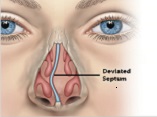
Background: The primary aim of this study was to compare palate depth and nasal septum deviation between patients with unilateral and bilateral buccal and palatal impactions of maxillary canines and those without impaction. Materials and Method: This cross-sectional study examined CBCT images of 60 patients from a private radiology archive, divided into four subgroups of 10 patients each with unilateral or bilateral buccal and palatal impactions, and a control group of 20 patients without impaction. Nasal deviation was assessed by measuring the distance of the maximum convexity of the deviated septum from the midsagittal plane in the coronal CBCT cut. Palate depth (PD) was measured as the perpendicular line from the middle of the axis connecting the mesiopalatal cusp of the first molar to the hard palate. Measurements were performed using NNT 16.3.1 software and validated by radiology and orthodontics specialists. Independent t-tests were used for statistical comparisons. Results: There were no significant differences in palate depth (p > 0.05) or nasal septum deviation (p > 0.05) between the control group and patients with unilateral or bilateral buccal and palatal impactions of maxillary canines. Conclusion: The study found no significant differences in palate depth and nasal septum deviation between patients with and without maxillary canine impaction, suggesting that impaction does not significantly affect these anatomical features. Further research is recommended to explore these findings in larger populations.
References
Alqerban A. Impacted maxillary canine in unilateral cleft lip and palate: A literature review. The Saudi dental journal. 2019 Jan 1;31(1):84-92.
https://doi.org/10.1016/j.sdentj.2018.11.001
PMid:30705572 PMCid:PMC6349903
Litsas G, Acar A. A review of early displaced maxillary canines: etiology, diagnosis and interceptive treatment. The open dentistry journal. 2011 Mar 16;5:39.
https://doi.org/10.2174/1874210601105010039
PMid:21566691 PMCid:PMC3091288
Becker A, Chaushu G, Chaushu S. Analysis of failure in the treatment of impacted maxillary canines. American Journal of Orthodontics and Dentofacial Orthopedics. 2010 Jun 1;137(6):743-54.
https://doi.org/10.1016/j.ajodo.2008.07.022
PMid:20685529
Fattahi HR, Pakshir HR. Use of panoramic radiographs in localization of maxillary impacted canines. Journal of Dentistry. 2005 Dec 1;6(3, 4):65-72.
D' Oleo-Aracena MF, Arriola-Guillén LE, Rodríguez-Cárdenas YA, Ruíz-Mora GA. Skeletal and dentoalveolar bilateral dimensions in unilateral palatally impacted canine using cone beam computed tomography. Progress in orthodontics. 2017 Dec;18:1-7.
https://doi.org/10.1186/s40510-017-0160-6
PMid:28164257 PMCid:PMC5316518
Momeni Danaie S, Salehi P, Kalantary MH. The importance of maxillary canine: A review. Journal of Dentistry. 2019 Jan 22;4(3):53-61.
Liuk W, Olive RJ, Griffin M, Monsour P. Associations between palatally displaced canines and maxillary lateral incisors. American Journal of Orthodontics and Dentofacial Orthopedics. 2013 May 1;143(5):622-32.
https://doi.org/10.1016/j.ajodo.2012.11.025
PMid:23631964
Kanavakis G, Curran KM, Wiseman KC, Barone NP, Finkelman MD, Srinivasan S, Lee MB, Trotman CA. Evaluation of crown-root angulation of lateral incisors adjacent to palatally impacted canines. Progress in Orthodontics. 2015 Dec;16:1-6.
https://doi.org/10.1186/s40510-015-0074-0
PMid:25749110 PMCid:PMC4385030
Cruz RM. Orthodontic traction of impacted canines: Concepts and clinical application. Dental press journal of orthodontics. 2019 Jan;24:74-87.
https://doi.org/10.1590/2177-6709.24.1.074-087.bbo
PMid:30916252 PMCid:PMC6434671
Kajan ZD, Khademi J, Nemati S, Niksolat E. The effects of septal deviation, concha bullosa, and their combination on the depth of posterior palatal arch in cone-beam computed tomography. Journal of Dentistry. 2016 Mar;17(1):26.
Peck S. The palatally displaced canine as a dental anomaly of genetic origin. Angle Orthod. 1995;65:95-102.
Sharma S, Sharma P, Rathore A, Raza M. 3-D assessment of skeletal and dentoalveolar bilateral dimensions in unilateral impacted palatal canine cases-A CBCT study. Journal of Clinical and Experimental Dentistry. 2023 Dec 1;15(12):e1007.
https://doi.org/10.4317/jced.60982
PMid:38186918 PMCid:PMC10767739
Yassaei S, Safi Y, Valian F, Mohammadi A. Evaluation of maxillary arch width and palatal volume and depth in patients with maxillary impacted canine by CBCT. Heliyon. 2022 Oct 3;8(10):e10854.
https://doi.org/10.1016/j.heliyon.2022.e10854
PMid:36247115 PMCid:PMC9561739
Kareem FA, Rasheed TA, Rauf AM, Jalal RA, Faraj BM. Three-dimensional measurements of the Palate and Dental Arch Perimeter as predictors for Maxillary Palatal Canine Impaction-A Cone-Beam Computed Tomography Image Analysis. Diagnostics. 2023 May 20;13(10):1808.
https://doi.org/10.3390/diagnostics13101808
PMid:37238292 PMCid:PMC10217248
Schindel RH, Duffy SL. Maxillary transverse discrepancies and potentially impacted maxillary canines in mixed-dentition patients. The Angle Orthodontist. 2007 May 1;77(3):430-5.
https://doi.org/10.2319/0003-3219(2007)077[0430:MTDAPI]2.0.CO;2
PMid:17465649
Elmarhoumy SM, Safwat W, Ellaithy M. Evaluation of Palatal Depth and Nasal Septum in Patients with Unilaterally Impacted Maxillary Canines: A Cone Beam Tomography Study. Al-Azhar Journal of Dental Science. 2023 Apr 1;26(2):241-6.
https://doi.org/10.21608/ajdsm.2023.185329.1404
Sharhan HM, Almashraqi AA, Al-Fakeh H, Alhashimi N, Abdulghani EA, Chen W, Al-Sosowa AA, Cao B, Alhammadi MS. Qualitative and quantitative three-dimensional evaluation of maxillary basal and dentoalveolar dimensions in patients with and without maxillary impacted canines. Progress in Orthodontics. 2022 Oct 24;23(1):38.
https://doi.org/10.1186/s40510-022-00434-3
PMid:36274114 PMCid:PMC9588850
Sobhani F, Miresmaeili A, Mahjub H, Farhadian M. Statistical shape analysis of maxillary palatal morphology in patients with palatally displaced canines. BMC Medical Imaging. 2023 Nov 29;23(1):198.
https://doi.org/10.1186/s12880-023-01158-4
PMid:38031064 PMCid:PMC10685537
Genc E, Karaman A. Investigation of the relationship between maxillary dimensions and labial and palatal maxillary impacted canines using cone beam computed tomography. Journal of Stomatology, Oral and Maxillofacial Surgery. 2023 Feb 1;124(1):101282.
https://doi.org/10.1016/j.jormas.2022.09.003
PMid:36087915
Erhamza TS, Akan B. Is there a relationship between buccally displaced maxillary canine and nasal septum deviation?. Eastern Journal of Medicine. 2021;26(1):53-6.
https://doi.org/10.5505/ejm.2021.77698
Tassoker M, Magat G, Lale B, Gulec M, Ozcan S, Orhan K. Is the maxillary sinus volume affected by concha bullosa, nasal septal deviation, and impacted teeth A CBCT study. European Archives of Oto-Rhino-Laryngology. 2020 Jan;277:227-33.
https://doi.org/10.1007/s00405-019-05651-x
PMid:31542830
Kucukkaraca E. Is There a Relationship Between Unilateral/Bilateral Impacted Maxillary Canines and Nasal Septum Deviation? Cureus. 2023 Oct 29;15(10):e47931.
https://doi.org/10.7759/cureus.47931
PMid:38034237 PMCid:PMC10684973
Yassaei S, Safi Y, Valian F, Mohammadi A. Evaluation of maxillary arch width and palatal volume and depth in patients with maxillary impacted canine by CBCT. Heliyon. 2022 Oct 3;8(10):e10854.
https://doi.org/10.1016/j.heliyon.2022.e10854
PMid:36247115 PMCid:PMC9561739
Farhadifard H, Shokri A, Salehzadeh M, Farhadian M, Ahmadpour Y. Evaluation of the relationship between maxillary canine impaction with arch dimensions and maxillary sinus dimensions using Cone Beam Computed Tomography (CBCT). World Journal of Plastic Surgery. 2024;13(2):32.
https://doi.org/10.61186/wjps.13.2.32
PMid:39193243 PMCid:PMC11346695
Fattahi H, Ghaeed F, Alipour A. Association between maxillary canine impaction and arch dimensions. Australasian Orthodontic Journal. 2012 May 1;28(1):57-62.
Published
How to Cite
Issue
Section
License
Copyright (c) 2024 Galen Medical Journal

This work is licensed under a Creative Commons Attribution 4.0 International License.


