Comprehensive Assessment of Anterior and Posterior Lingual Mandibular Depressions Using Cone Beam Computed Tomography: Morphology, Prevalence and Clinical Implications
DOI:
https://doi.org/10.31661/gmj.v13iSP1.3703Keywords:
Cone-Beam Computed Tomography; Dental implants; Lingual subfossa; MandibleAbstract
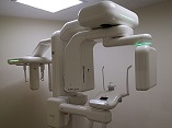
Background: Assessment of lingual mandibular depressions, has significant clinical relevance in various maxillofacial procedures. Cone Beam Computed Tomography (CBCT) has emerged as a valuable tool for the detailed evaluation of these anatomical features. The aim of this study was to comprehensively assess lingual mandibular depressions utilizing CBCT imaging. Materials and Methods: In this descriptive-cross-sectional study, 384 images from patients were examined. The images were reviewed using the Plunmeca Promax 3D device. the concavity depth, ridge thickness from the alveolar crest area, angle of concavity two millimeters above the inferior alveolar nerve, height of concavity from the start of the concavity to its end, and also the linear height along the occlusal plane with the opposing teeth in the lower jaw ridge were measured in the lingual area of the canine-premolar. Results: The frequency of concavity was 2.9% in the canine-premolar region, in the first molar region 34.7%, and in the second molar region 98.2%. The concavity depth in the canine-premolar region was measured at 4.41 millimeters, in the first molar region at 3.80 millimeters, and the second molar region at 4.43 millimeters. The concavity height was reported as 13.26 millimeters in the canine-premolar region, 12.35 millimeters in the first molar region, and 13.51 millimeters in the second molar region. The angle of concavity was measured at 60.48 degrees in the canine-premolar region. Ridge thickness in the canine-premolar region was 9.06 millimeters, in the first molar region 10.47 millimeters, and the second molar region 10.43 millimeters. No interference was found in the canine-premolar region, while interference was observed in 7.25% of cases in the first molar region and 23.6% in the second molar region. Conclusion: Imaging with CBCT should be performed before implant placement also the concavity depth in the area should be considered to avoid potential interference during implant placement.
References
Pires FR, Bruzigueses Espíndola CB, Ferreira Espíndola SH, Netto JNS. Anterior lingual mandibular bone depression: differential diagnosis of periapical inflammatory disorders. Gen Dent. 2018;66(5):e6-e11.
Friedrich RE, Barsukov E, Kohlrusch FK, Zustin J, Hagel C, Speth U, et al. Lingual mandibular bone depression. in vivo. 2020;34(5):2527-41.
https://doi.org/10.21873/invivo.12070
PMid:32871782 PMCid:PMC7652515
Haghanifar S, Arbabzadegan N, Moudi E, Bijani A, Nozari F. Evaluation of lingual mandibular depression of the submandibular salivary glands using cone-beam computed tomography. Res Med Dent Sci. 2018;6:563-7.
Philipsen H, Takata T, Reichart P, Sato S, Suei Y. Lingual and buccal mandibular bone depressions: a review based on 583 cases from a world-wide literature survey, including 69 new cases from Japan. Dentomaxillofacia Radiol. 2002;31(5):281-90.
https://doi.org/10.1038/sj.dmfr.4600718
PMid:12203126
Liu L, Kang BC, Yoon SJ, Lee JS, Hwang SA. Radiographic features of lingual mandibular bone depression using dental cone beam computed tomography. Dentomaxillofacia Radiol. 2018;47(6):20170383.
https://doi.org/10.1259/dmfr.20170383
PMid:29589968 PMCid:PMC6196052
Zonouz AT, Azar FP, Pourzare S, Hosinifard H, Molaei Z. Association between dental amalgam fillings and multiple sclerosis: A systematic review and meta-analysis. J Res Clin Med. 2023;11(1):18.
https://doi.org/10.34172/jrcm.2023.31924
Al-Sadhan Re. CT study of the medial depression of the human mandibular ramus Int. J Morphol. 2021;39(6):1570-4.
Hasani M, Karandish M, Salari Y. Characteristics of Medial Depression of the Mandibular Ramus: A CBCT Analysis in Different Sagittal Skeletal Patterns. J Dent Shiraz Univ Med Sci. 2023;24(1):1.
Monteiro LS, Da Câmara MI, Tadeu F, Salazar F, Pacheco JJ. Posterior lingual bone depression diagnosis using 3D-computed tomography. Revista Portuguesa de Estomatologia, Medicina Dentária e Cirurgia Maxilofacial. 2012 Jul 1;53(3):170-4.
https://doi.org/10.1016/j.rpemd.2012.05.007
Altwaim M, Al-Sadhan R. Bilateral Anterior Lingual Depression in the Mandible: Cone Beam Computed Tomography Case Report and Review of the Literature. Cureus. 2019;11(12):e6348.
https://doi.org/10.7759/cureus.6348
PMid:31886090 PMCid:PMC6907715
Peñarrocha-Diago M, Balaguer-Martí JC, Peñarrocha-Oltra D, Bagán J, Peñarrocha-Diago M, Flanagan D. Floor of the mouth hemorrhage subsequent to dental implant placement in the anterior mandible. Clin Cosmet Investig Dent. 2019:235-42.
https://doi.org/10.2147/CCIDE.S207120
PMid:31496828 PMCid:PMC6690045
Alshenaiber R, Barclay C, Silikas N. The Effect of Mini Dental Implant Number on Mandibular Overdenture Retention and Attachment Wear. BioMed Research International. 2023;2023(1):7099761.
https://doi.org/10.1155/2023/7099761
PMid:37168235 PMCid:PMC10164865
Simon F, Wolfe SA, Guichard B, Bertolus C, Khonsari RH. Paul Tessier facial reconstruction in 1970 Iran, a series of post-noma defects. Journal of Craniomaxillofac Surg. 2015;43(4):503-9.
https://doi.org/10.1016/j.jcms.2015.02.014
PMid:25817742
Bressan E, Paniz G, Lops D, Corazza B, Romeo E, Favero G. Influence of abutment material on the gingival color of implant‐supported all‐ceramic restorations: a prospective multicenter study. Clin Oral Implants Res. 2011;22(6):631-7.
https://doi.org/10.1111/j.1600-0501.2010.02008.x
PMid:21070378
Panjnoush M, Eil N, Kheirandish Y, Mofidi N, Shamshiri AR. Evaluation of the concavity depth and inclination in jaws using CBCT. Caspian J of Dent Res. 2016;5(2):17-23.
Herranz-Aparicio J, Marques J, Almendros-Marqués N, Gay-Escoda C. Retrospective study of the bone morphology in the posterior mandibular region, Evaluation of the prevalence and the degree of lingual concavity and their possible complications. Med Oral Patol Oral Cir Bucal. 2016 Oct 1;21(6):e731.
https://doi.org/10.4317/medoral.21256
PMid:27694785 PMCid:PMC5116115
Liu L, Kang BC, Yoon SJ, Lee JS, Hwang SA. Radiographic features of lingual mandibular bone depression using dental cone beam computed tomography. Dentomaxillofac Radiol. 2018 Jul 1;47(6):20170383.
https://doi.org/10.1259/dmfr.20170383
PMid:29589968 PMCid:PMC6196052
Nickenig H-J, Wichmann M, Eitner S, Zoeller JE, Kreppel M. Lingual concavities in the mandible: a morphological study using cross-sectional analysis determined by CBCT. J CRANIO MAXILL SURG. 2015;43(2):254-9.
https://doi.org/10.1016/j.jcms.2014.11.018
PMid:25547216
Chan HL, Brooks SL, Fu JH, Yeh CY, Rudek I, Wang HL. Cross‐sectional analysis of the mandibular lingual concavity using cone beam computed tomography. Clin Oral Implants Res. 2011;22(2):201-6.
https://doi.org/10.1111/j.1600-0501.2010.02018.x
PMid:21044167

Published
How to Cite
Issue
Section
License
Copyright (c) 2024 Galen Medical Journal

This work is licensed under a Creative Commons Attribution 4.0 International License.


