
In Vitro Comparative Accuracy of PSP Digital Radiography and CBCT for Detection of Broken Nickel-Titanium Files in Endodontically Treated Root Canals
DOI:
https://doi.org/10.31661/gmj.v13iSP1.3711Keywords:
Root Canal Therapy; Cone-Beam Computed Tomography; Radiography, Dental, Digital; Nickel-TitaniumAbstract
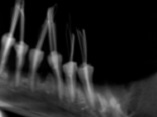
Background: This study aimed to compare the accuracy of indirect photostimulable phosphor (PSP) digital radiography and cone-beam computed tomography (CBCT) for detection of broken nickel-titanium (NiTi) files in endodontically treated root canals. Materials and Methods: This in vitro study was conducted on 108 extracted single-rooted mandibular premolars in 4 group (n=27) of positive control (root canal instrumentation and obturation), negative control (root canal instrumentation without obturation) and two experimental groups of file fracture with and without root canal obturation. The teeth underwent PSP digital radiography and CBCT, and the radiographs were evaluated by one oral radiologist and one endodontist twice. Presence/absence of a broken file in the root canals was reported using a 4-point scale. The sensitivity, specificity, and accuracy were calculated and compared for CBCT and PSP digital radiography. Results: The sensitivity, specificity, and accuracy for detection of broken NiTi files in obturated canals were 51.9%, 59.3% and 55.6%, respectively for CBCT and 70.3%, 85%, and 77.8%, respectively for PSP radiography. These values were 81.4%, 59.3%, and 79.6%, respectively for CBCT and 85.1%, 81.4%, and 83.3%, respectively for PSP radiography in unfilled canals. PSP digital radiography was significantly superior to CBCT for detection of broken files in obturated (P=0.01) but not in unfilled (P=0.420) root canals. Conclusion: Considering the lower radiation dose and higher accuracy of PSP digital radiography than CBCT for detection of broken NiTi files in filled canals, and their comparable accuracy in unfilled canals, PSP digital radiography is recommended for this purpose.
References
Lin LM, Rosenberg PA, Lin J. Do procedural errors cause endodontic treatment failure?. The Journal of the American Dental Association. 2005 Feb 1;136(2):187-93.
https://doi.org/10.14219/jada.archive.2005.0140
PMid:15782522
Shen Y, Coil JM, McLean AG, Hemerling DL, Haapasalo M. Defects in nickel-titanium instruments after clinical use. Part 5: single use fromendodontic specialty practices. J Endod 2009; 35: 1363-7.
https://doi.org/10.1016/j.joen.2009.07.004
PMid:19801231
Sattapan B, Nervo GJ, Palamara JE, Messer HH. Defects in rotary nickel-titanium files after clinical use. Journal of endodontics. 2000 Mar 1;26(3):161-5.
https://doi.org/10.1097/00004770-200003000-00008
PMid:11199711
Saunders JL, Eleazer PD, Zhang P, Michalek S. Effect of a separated instrument on bacterial penetration of obturated root canals. Journal of endodontics. 2004 Mar 1;30(3):177-9.
https://doi.org/10.1097/00004770-200403000-00012
PMid:15055438
Di Fiore PM, Genov KA, Komaroff E, Li Y, Lin L. Nickel-titanium rotary instrument fracture: a clinical practice assessment. International Endodontic Journal. 2006 Sep;39(9):700-8.
https://doi.org/10.1111/j.1365-2591.2006.01137.x
PMid:16916359
Hargreaves Km , Cohen S , Berman lh. Cohen's pathways of the pulp. 10th ed: moseby inc; 2011.
Kleier DJ, Shibilski K, Averbach RE. Radiographic appearance of titanium posts in endodontically treated teeth. Journal of Endodontics. 1999 Feb 1;25(2):128-31.
https://doi.org/10.1016/S0099-2399(99)80012-0
PMid:10204472
Givol N, Rosen E, Taicher S, Tsesis I. Risk management in endodontics. Journal of endodontics. 2010 Jun 1;36(6):982-4.
https://doi.org/10.1016/j.joen.2010.03.030
PMid:20478450
Bahcall JK. Remedying and preventing endodontic rotary nickel-titanium (NiTi) file breakage. Compendium of Continuing Education in Dentistry . 2013 May 1;34(5): 15488578.
Parashos P, Messer HH. Rotary NiTi instrument fracture and its consequences. Journal of endodontics. 2006 Nov 1;32(11):1031-43.
https://doi.org/10.1016/j.joen.2006.06.008
PMid:17055902
Algarni Y. Fracture Incidence of New Reciprocating Nickel-Titanium (NiTi) Files: A Cross-Sectional Retrospective Study. Cureus. 2024 Aug 25;16(8):e67762.
https://doi.org/10.7759/cureus.67762
Reit C, Petersson K , Molven O. Diagnosis of Pulpal and Periradicular Disease. Textbook of Endodontology, Blackwell Publishing Ltd: Oxford, UK, 1st edition; 2003.
Scarfe WC, Levin MD, Gane D, Farman AG. Use of cone beam computed tomography in endodontics. International journal of dentistry. 2009;2009(1):634567.
https://doi.org/10.1155/2009/634567
PMid:20379362 PMCid:PMC2850139
White SC, Pharoah MJ. Oral Radiology principles and interprentation. 8th ed: chaper 4 page 40 ; 2019 .
Eskandarloo A, Mirshekari A, Poorolajal J, Mohammadi Z, Shokri A. Comparison of cone-beam computed tomography with intraoral photostimulable phosphor imaging plate for diagnosis of endodontic complications: a simulation study. Oral surgery, oral medicine, oral pathology and oral radiology. 2012 Dec 1;114(6):e54-61.
https://doi.org/10.1016/j.oooo.2012.05.026
PMid:22981099
Shemesh H, Cristescu RC, Wesselink PR, Wu MK. The use of cone-beam computed tomography and digital periapical radiographs to diagnose root perforations. Journal of endodontics. 2011 Apr 1;37(4):513-6.
https://doi.org/10.1016/j.joen.2010.12.003
PMid:21419300
Jeffcoat MK, Wang IC, Reddy MS. Radiographic diagnosis in periodontics. Periodontology 2000. 1995 Feb;7(1):54-68.
https://doi.org/10.1111/j.1600-0757.1995.tb00036.x
PMid:9567930
Kamburoğlu KI, Kursun S. A comparison of the diagnostic accuracy of CBCT images of different voxel resolutions used to detect simulated small internal resorption cavities. International endodontic journal. 2010 Sep;43(9):798-807.
https://doi.org/10.1111/j.1365-2591.2010.01749.x
PMid:20609023
Tyndall DA, Rathore S. Cone-beam CT diagnostic applications: caries, periodontal bone assessment, and endodontic applications. Dental Clinics of North America. 2008 Oct 1;52(4):825-41.
https://doi.org/10.1016/j.cden.2008.05.002
PMid:18805231
D'addazio PS, Campos CN, Özcan M, Teixeira HG, Passoni RM, Carvalho AC. A comparative study between cone-beam computed tomography and periapical radiographs in the diagnosis of simulated endodontic complications. International endodontic journal. 2011 Mar;44(3):218-24.
https://doi.org/10.1111/j.1365-2591.2010.01802.x
PMid:21039626
Bushong SC. Radiologic Science for technologists: physics, biology, and protection. 9th ed. Canada:St. louis: Mosby Elsevier; 2008. P.273-93.
Ayatollahi F, Tabrizizadeh M, Razavi H, Mowji M. Diagnostic value of cone-beam computed tomography and digital periapical radiography in detection of separated instruments. Iranian Endodontic Journal. 2019;14(1):14.
Brito AC, Verner FS, Junqueira RB, Yamasaki MC, Queiroz PM, Freitas DQ, Oliveira-Santos C. Detection of fractured endodontic instruments in root canals: comparison between different digital radiography systems and cone-beam computed tomography. Journal of endodontics. 2017 Apr 1;43(4):544-9.
https://doi.org/10.1016/j.joen.2016.11.017
PMid:28216273
Madian S, Gaweesh Y, El-Badawy F, Genena S. Diagnostic efficacy of 3 imaging modalities in the detection of fractured endodontic instruments: an in vitro study. Oral Surgery, Oral Medicine, Oral Pathology and Oral Radiology. 2023 Feb 1;135(2):303-11.
https://doi.org/10.1016/j.oooo.2022.09.012
Alemam S, Abuelsadat SH , Saber SH ,Elsewify T. Accuracy, sensitivity and specificity of three imaging modalities in detection of separated intracanal Instrument. BUE scholar. 2020;34: (97-103).
Abdinian M, Moshkforoush S, Hemati H, Soltani P, Moshkforoushan M, Spagnuolo G. Comparison of cone beam computed tomography and digital radiography in detecting separated endodontic files and strip perforation. Applied Sciences. 2020 Dec 5;10(23):8726.
https://doi.org/10.3390/app10238726
D'addazio PS, Campos CN, Özcan M, Teixeira HG, Passoni RM, Carvalho AC. A comparative study between cone-beam computed tomography and periapical radiographs in the diagnosis of simulated endodontic complications. International endodontic journal. 2011 Mar;44(3):218-24.
https://doi.org/10.1111/j.1365-2591.2010.01802.x
PMid:21039626
Haghanifar S, Moudi E, Mesgarani A, Bijani A, Abbaszadeh N. A comparative study of cone-beam computed tomography and digital periapical radiography in detecting mandibular molars root perforations. Imaging science in dentistry. 2014 Jun 1;44(2):115-9.
https://doi.org/10.5624/isd.2014.44.2.115
PMid:24944960 PMCid:PMC4061294
Rosen E, Venezia NB, Azizi H, Kamburoglu K, Meirowitz A, Ziv-Baran T, Tsesis I. A comparison of cone-beam computed tomography with periapical radiography in the detection of separated instruments retained in the apical third of root canal-filled teeth. Journal of endodontics. 2016 Jul 1;42(7):1035-9.
https://doi.org/10.1016/j.joen.2016.04.016
PMid:27238414
Talwar S, Utneja S, Nawal RR, Kaushik A, Srivastava D, Oberoy SS. Role of cone-beam computed tomography in diagnosis of vertical root fractures: a systematic review and meta-analysis. Journal of Endodontics. 2016 Jan 1;42(1):12-24.
https://doi.org/10.1016/j.joen.2015.09.012
PMid:26699923
Dutra KL, Haas L, Porporatti AL, Flores-Mir C, Santos JN, Mezzomo LA, Correa M, Canto GD. Diagnostic accuracy of cone-beam computed tomography and conventional radiography on apical periodontitis: a systematic review and meta-analysis. Journal of endodontics. 2016 Mar 1;42(3):356-64.
https://doi.org/10.1016/j.joen.2015.12.015
PMid:26902914
Elsaltani MH, Farid MM, Ashmawy MS. Detection of simulated vertical root fractures: which cone-beam computed tomographic system is the most accurate?. Journal of Endodontics. 2016 Jun 1;42(6):972-7.
https://doi.org/10.1016/j.joen.2016.03.013
PMid:27130336
Chang E, Lam E, Shah P, Azarpazhooh A. Cone-beam computed tomography for detecting vertical root fractures in endodontically treated teeth: a systematic review. Journal of endodontics. 2016 Feb 1;42(2):177-85.
https://doi.org/10.1016/j.joen.2015.10.005
PMid:26631300
Ardakani FE, Razavi SH, Tabrizizadeh M. Diagnostic value of cone-beam computed tomography and periapical radiography in detection of vertical root fracture. Iranian endodontic journal. 2015;10(2):122.
Published
How to Cite
Issue
Section
License
Copyright (c) 2024 Galen Medical Journal

This work is licensed under a Creative Commons Attribution 4.0 International License.


