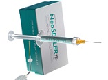
Tissue Reaction Elicited by Neosealer Flo, AH 26, and CC Sealer in Rats
DOI:
https://doi.org/10.31661/gmj.v13iSP1.3599Keywords:
Animal; Canals Sealer; Biocompatible Materials; Calcium SilicateAbstract
Background: Since root canal sealers are in contact with periradicular tissues, biocompatibility is one of their most important features .There is no available study about the biocompatibility of NSF and CC Sealer that are newly made bioceramic-based sealers. This study aimed to compare the tissue reaction elicited by NeoSealer Flo (NSF), AH26, and ColdCeramic Sealer (CC sealer) in rats. Material and Methods: The sealers were mixed and applied in molds to fabricate sealer discs, which were then implanted in the subcutaneous tissue of the backs of 30 healthy adult Albino Wistar rats. Each rat received three sealer discs and the fourth incision site remained empty as a control group. After 7, 30, and 90 days, the rats were sacrificed. Biopsy samples were evaluated regarding the extent and severity of inflammation, angiogenesis, fibroplasia, and infiltration. Data were analyzed in SPSS software using Kruskal-Wallis and Mann-Whitney U tests. Results: Tissue reaction to NSF was generally severe and increased up to day 30, but slightly decreased at three months, although it was still severe, and significantly greater than the tissue reaction to other sealer types. After one month, all rats in the NSF group showed foreign body reaction and giant cells around sealer particles; while, foreign body reaction was not seen in other groups. Tissue reaction to CC Sealer and AH26 was not significantly different at any point in time (P>0.05) and was the highest on day seven and then decreased up to month three. Conclusion: According to the present results, the CC Sealer appears to be a biocompatible material; however, NSF showed higher severity and extent of inflammation and triggered higher tissue reaction.
References
Torabinejad M, Ashraf F, Shabahang sh . Endodontics: principles and practice. Elsevier Health Sciences. (2021); :327-334.
Haji T H, Selivany B J, Suliman A A. Sealing ability in vitro study and biocompatibility in vivo animal study of different bioceramic based sealers. Clinical and experimental dental research. (2022); 8(6): 1582-1590.
https://doi.org/10.1002/cre2.652
PMid:36397655 PMCid:PMC9760163
Saxena P, Gupta S K, Newaskar V. Biocompatibility of root-end filling materials: recent update. Restorative dentistry & endodontics. (2013); 38(3): 119-127.
https://doi.org/10.5395/rde.2013.38.3.119
PMid:24010077 PMCid:PMC3761119
Al-Haddad A, Che Ab Aziz Z A . Bioceramic-Based Root Canal Sealers: A Review. International journal of biomaterials. (2016); 2016: 9753210.
https://doi.org/10.1155/2016/9753210
PMid:27242904 PMCid:PMC4868912
Kishen A, Peters O A, Zehnder M, Diogenes A R, Nair M K . Advances in endodontics: Potential applications in clinical practice. Journal of conservative dentistry : JCD. (2016); 19(3): 199-206.
https://doi.org/10.4103/0972-0707.181925
PMid:27217630 PMCid:PMC4872571
Yang X, Zheng T, Yang N, Yin Z, Wang W, Bai Y . A Review of the research methods and progress of biocompatibility evaluation of root canal sealers. Australian endodontic journal : the journal of the Australian Society of Endodontology Inc. (2023); 49(1): 508-514.
https://doi.org/10.1111/aej.12725
PMid:36480411
de Oliveira R L, Oliveira Filho R S, Gomes HdeC, de Franco M F, Enokihara M M, Duarte M A . Influence of calcium hydroxide addition to AH Plus sealer on its biocompatibility. Oral surgery, oral medicine, oral pathology, oral radiology, and endodontics. (2010); 109(1): e50-e54.
https://doi.org/10.1016/j.tripleo.2009.08.026
PMid:20123371
Modaresi J, Talakoob M H . Comparison of two root-end filling materials . Journal of Isfahan Dental School. (2015);11(5): 379- 386.
Modaresi J, Yavari S A, Dianat S O, Shahrabi S . A comparison of tissue reaction to MTA and an experimental root-end restorative material in rats. Australian endodontic journal : the journal of the Australian Society of Endodontology Inc. (2005); 31(2): 69-72.
https://doi.org/10.1111/j.1747-4477.2005.tb00229.x
PMid:16128256
Zamparini F, Prati C, Taddei P, Spinelli A, Di Foggia M, Gandolfi M G . Chemical-Physical Properties and Bioactivity of New Premixed Calcium Silicate-Bioceramic Root Canal Sealers. International journal of molecular sciences. (2022); 23(22): 13914.
https://doi.org/10.3390/ijms232213914
PMid:36430393
Basta D G, Reslan M R, Rayyan M, Sayed M . Evaluation of Antibacterial Effect of New Sealer "Neoseal" and Two Commercially Used Endodontic Sealers against Enterococcus faecalis: An In Vitro Study. The journal of contemporary dental practice. (2023); 24(11): 871-876.
https://doi.org/10.5005/jp-journals-10024-3599
PMid:38238275
Bernáth M, Szabó J . Tissue reaction initiated by different sealers. International endodontic journal. (2003); 36(4): 256-261.
https://doi.org/10.1046/j.1365-2591.2003.00662.x
PMid:12702119
Paul Flecknell . Laboratory Animal Anaesthesia, Newcastle-upon-Tyne, UK. Elsevier Health Sciences. (2009); :187.
https://doi.org/10.1016/B978-0-12-369376-1.00002-2
Assmann E, Böttcher D E, Hoppe C B, Grecca FS, Kopper P M . Evaluation of bone tissue response to a sealer containing mineral trioxide aggregate. Journal of endodontics. (2015); 41(1): 62-66.
https://doi.org/10.1016/j.joen.2014.09.019
PMid:25447498
Marjani M, Hashemi M, Sedaghat R . Histopathologic comparison of chromic catgut suture materials from Iran and abroad. Journal of Mazandaran University of Medical Sciences. (2010); 19(74): 33-42.
Silva E C A, Pradelli J A, da Silva G F, Cerri P S, Tanomaru-Filho M, Guerreiro-Tanomaru J M . Biocompatibility and bioactive potential of NeoPUTTY calcium silicate-based cement: An in vivo study in rats. International endodontic journal. (2024); 57(6): 713-726.
https://doi.org/10.1111/iej.14054
PMid:38467586
Tavares C O, Böttcher D E, Assmann E, Kopper P M, de Figueiredo J A, Grecca F S, Scarparo R K . Tissue reactions to a new mineral trioxide aggregate-containing endodontic sealer. Journal of endodontics. (2013); 39(5): 653-657.
https://doi.org/10.1016/j.joen.2012.10.009
PMid:23611385
Ashraf H, Shafagh P, Mashhadi Abbas F, Heidari S, Shahoon H, Zandian A, Aghajanpour L, Zadsirjan S . Biocompatibility of an experimental endodontic sealer (Resil) in comparison with AH26 and AH-Plus in rats: An animal study. Journal of dental research, dental clinics, dental prospects. (2022); 16(2): 112-117.
https://doi.org/10.34172/joddd.2022.019
PMid:36561386 PMCid:PMC9763656
Kohsar A H, Hasani M, Karami M, Moosazadeh M, Dashti A, Shiva A . Subcutaneous Tissue Response to Adseal and Sure-Seal Root Sealers in Rats: a Histopathological Study. Maedica. (2022); 17(3): 654-661.
https://doi.org/10.26574/maedica.2022.17.3.654
PMid:36540595 PMCid:PMC9720645
Hoshino R A, Delfino M M, da Silva G F, Guerreiro-Tanomaru J M, Tanomaru-Filho M, Sasso-Cerri E, Cerri P S . Biocompatibility and bioactive potential of the NeoMTA Plus endodontic bioceramic-based sealer. Restorative dentistry & endodontics. (2020); 46(1): e4.
https://doi.org/10.5395/rde.2021.46.e4
PMid:33680893 PMCid:PMC7906839
Feghali C A, Wright T M . Cytokines in acute and chronic inflammation. Frontiers in bioscience : a journal and virtual library. (1997); 2: d12-d26.
PMid:9159205
Cardinali F, Camilleri J . A critical review of the material properties guiding the clinician's choice of root canal sealers. Clinical oral investigations. (2023); 27(8): 4147-4155.
https://doi.org/10.1007/s00784-023-05140-w
PMid:37460901 PMCid:PMC10415471
Mozayeni M A, Salem Milani A, Alim Marvasti L, Mashadi Abbas F, Modaresi S J . Cytotoxicity of Cold Ceramic Compared with MTA and IRM. Iranian endodontic journal. (2009); 4(3):106-111.
Figueiredo J A, Pesce H F, Gioso M A, Figueiredo M A. The histological effects of four endodontic sealers implanted in the oral mucosa: submucous injection versus implant in polyethylene tubes. International endodontic journal. (2001); 34(5): 377-385.
https://doi.org/10.1046/j.1365-2591.2001.00407.x
PMid:11482721

Published
How to Cite
Issue
Section
License
Copyright (c) 2024 Galen Medical Journal

This work is licensed under a Creative Commons Attribution 4.0 International License.


