
Comparison of the Accuracy of CBCT and MDCT Images in Measuring the Thickness of the Posterior Footplate of the Middle Ear in Iranian
DOI:
https://doi.org/10.31661/gmj.vi.3900Keywords:
Middle Ear; Footplate Thickness; CBCT; MDCTAbstract
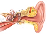
Background: Advancements in radiological imaging have transitioned from two-dimensional radiography to three-dimensional cone beam computed tomography (CBCT), offering high spatial resolution with reduced radiation doses compared to multidetector computed tomography (MDCT). While MDCT remains the standard for detailed visualization of bony structures like the ossicular chain, its higher radiation exposure is a concern. This study compares the accuracy of high-resolution (HR) and low-resolution (LR) CBCT with MDCT in measuring the thickness of the posterior footplate of the middle ear to identify a reliable, low-radiation alternative. Materials and Methods: Twelve adult human temporal bones from Imam Khomeini Hospital’s ENT Department were imaged using HR-CBCT, LR-CBCT (Jundishapur Dental School), and MDCT (Siemens Sensation 64-slice). Standardized imaging protocols ensured reproducibility, with measurements of posterior footplate thickness conducted in axial, coronal, and sagittal planes by two blinded radiologists. Data were analyzed using SPSS v23, with inter-modality agreement assessed via Kappa coefficient and differences evaluated with the McNemar test. Results: Mean posterior footplate thickness was 2.38 mm (HR-CBCT), 2.37 mm (LR-CBCT), and 2.23 mm (MDCT), with no significant differences (p > 0.05). HR-CBCT and LR-CBCT showed comparable accuracy to MDCT. Conclusion: CBCT, particularly HR-CBCT, offers a reliable, lower-radiation alternative to MDCT for otologic imaging, maintaining high resolution for middle ear assessments.
References
Robinson S, Suomalainen A, Kortesniemi M. mCT. MJEJOR. 2005;56(2):18591.
https://doi.org/10.1016/j.ejrad.2005.03.022
PMid:16233892
Sukovic P. Cone beam computed tomography in craniofacial imaging. Orthod Craniofac Res. 2003;6 Suppl 1:316; discussion 17982.
https://doi.org/10.1034/j.1600-0544.2003.259.x
PMid:14606532
Araki K, Maki K, Seki K, Sakamaki K, Harata Y, Sakaino R, et al. Characteristics of a newly developed dentomaxillofacial Xray cone beam CT scanner (CB MercuRay): system configuration and physical properties. Dentomaxillofac Radiol. 2004;33(1):519.
https://doi.org/10.1259/dmfr/54013049
PMid:15140823
Hashimoto K, Arai Y, Iwai K, Araki M, Kawashima S, Terakado M. A comparison of a new limited cone beam computed tomography apparatus for dental use with a multidetector row helical CT apparatus. Oral Surg Oral Med Oral Pathol Oral Radiol Endod. 2003;95(3):3717.
https://doi.org/10.1067/moe.2003.120
PMid:12627112
Maroldi R, Farina D, Palvarini L, Marconi A, Gadola E, Menni K, et al. Computed tomography and magnetic resonance imaging of pathologic conditions of the middle ear. Eur J Radiol. 2001;40(2):7893.
https://doi.org/10.1016/S0720-048X(01)00376-X
PMid:11704355
Dalchow CV, Weber AL, Yanagihara N, Bien S, Werner JA. Digital volume tomography: radiologic examinations of the temporal bone. AJR Am J Roentgenol. 2006;186(2):41623.
https://doi.org/10.2214/AJR.04.1353
PMid:16423947
Bromberg N, Brizuela M. Dental Cone Beam Computed Tomography. [Updated 2023 Apr 19] In: StatPearls [Internet] Treasure Island (FL) StatPearls Publishing; Available from: https://www.ncbi.nlm.nih.gov/books/NBK592390/
Raghavan M, Ong AA, Carr MM. Complications after craniofacial surgery: a review from 2012 to 2020. Cureus. 2025 Feb 6;17(2): e78625.
https://doi.org/10.7759/cureus.78625
Carlson ML, Driscoll CL, Gifford RH, Service GJ, Tombers NM, HughesBorst BJ, et al. Implications of minimizing trauma during conventional cochlear implantation. Otol Neurotol. 2011;32(6):9628.
https://doi.org/10.1097/MAO.0b013e3182204526
PMid:21659922 PMCid:PMC4127076
Verbist BM, Frijns JH, Geleijns J, van Buchem MA. Multisection CT as a valuable tool in the postoperative assessment of cochlear implant patients. AJNR Am J Neuroradiol. 2005;26(2):424-9.
Aschendorff A, Kubalek R, Hochmuth A, Bink A, Kurtz C, Lohnstein P, et al. Imaging procedures in cochlear implant patientsevaluation of different radiological techniques. Acta Otolaryngol Suppl. 2004(552):469.
https://doi.org/10.1080/03655230410017175
PMid:26942827
DahmaniCausse M, Marx M, Deguine O, Fraysse B, Lepage B, Escude B. Morphologic examination of the temporal bone by cone beam computed tomography: comparison with multislice helical computed tomography. Eur Ann Otorhinolaryngol Head Neck Dis. 2011;128(5):2305.
https://doi.org/10.1016/j.anorl.2011.02.016
PMid:22014530
Theunisse HJ, Joemai RM, Maal TJ, Geleijns J, Mylanus EA, Verbist BM. Conebeam CT versus multislice CT systems for postoperative imaging of cochlear implantationa phantom study on image quality and radiation exposure using human temporal bones. Otol Neurotol. 2015;36(4):5929.
https://doi.org/10.1097/MAO.0000000000000673
PMid:25420084
Bartling SH, Gupta R, Torkos A, Dullin C, Eckhardt G, Lenarz T, et al. Flatpanel volume computed tomography for cochlear implant electrode array examination in isolated temporal bone specimens. Otol Neurotol. 2006;27(4):4918.
https://doi.org/10.1097/00129492-200606000-00010
Erovic BM, Chan HH, Daly MJ, Pothier DD, Yu E, Coulson C, et al. Intraoperative conebeam computed tomography and multislice computed tomography in temporal bone imaging for surgical treatment. Otolaryngol Head Neck Surg. 2014;150(1):10714.
https://doi.org/10.1177/0194599813510862
PMid:24170658
Kemp P, Stralen JV, De Graaf P, Berkhout E, Horssen PV, Merkus P. ConeBeam CT Compared to MultiSlice CT for the Diagnostic Analysis of Conductive Hearing Loss: A Feasibility Study. J Int Adv Otol. 2020;16(2):2226.
https://doi.org/10.5152/iao.2020.5883
PMid:32784161 PMCid:PMC7419106
Suomalainen A, Kiljunen T, Kaser Y, Peltola J, Kortesniemi M. Dosimetry and image quality of four dental cone beam computed tomography scanners compared with multislice computed tomography scanners. Dentomaxillofac Radiol. 2009;38(6):36778.
https://doi.org/10.1259/dmfr/15779208
PMid:19700530
Tschauner S, Marterer R, Nagy E, Singer G, Riccabona M, Sorantin E. Experiences with image quality and radiation dose of cone beam computed tomography (CBCT) and multidetector computed tomography (MDCT) in pediatric extremity trauma. Skeletal Radiol. 2020;49(12):193949.
https://doi.org/10.1007/s00256-020-03506-9
PMid:32535775 PMCid:PMC7652807
Loubele M, Bogaerts R, Van Dijck E, Pauwels R, Vanheusden S, Suetens P, et al. Comparison between effective radiation dose of CBCT and MSCT scanners for dentomaxillofacial applications. Dentomaxillofac Radiol. 2009;71(3):4618.
https://doi.org/10.1016/j.ejrad.2008.06.002
PMid:18639404
Burck I, Schneider SV, Balster S, Lehn A, Yel I, Albrecht MH, et al. Radiohistologic comparison study of temporal bone specimens after cochlear implant electrode array insertion. is conebeam CT superior to MDCT. 2021;216(3):7528.
https://doi.org/10.2214/AJR.20.23157
PMid:33439050
Debeaupte M, Hermann R, Pialat JB, Martinon A, Truy E, Ltaief Boudrigua A. Cone beam versus multidetector computed tomography for detecting hearing loss. Eur Arch Otorhinolaryngol. 2019;276(2):31521.
https://doi.org/10.1007/s00405-018-5214-y
PMid:30467778
Shweel M, Amer MI, Elshamanhory AF. A comparative study of conebeam CT and multidetector CT in the preoperative assessment of odontogenic cysts and tumors. The Egyptian Journal of Radiology and Nuclear Medicine. 2013;44(1):2332.
https://doi.org/10.1016/j.ejrnm.2012.12.002
Komori M, Yanagihara N, Hyodo J, Miuchi S. Position of TORP on the stapes footplate assessed with cone beam computed tomography. Otol Neurotol. 2012;33(8):13536.
https://doi.org/10.1097/MAO.0b013e31826a5260
PMid:22975904
Zou J, Lahelma J, Arnisalo A, Pyykko I. Clinically relevant human temporal bone measurements using novel highresolution conebeam CT. J Otol. 2017;12(1):917.
https://doi.org/10.1016/j.joto.2017.01.002
PMid:29937832 PMCid:PMC6011811
Published
How to Cite
Issue
Section
License
Copyright (c) 2025 Galen Medical Journal

This work is licensed under a Creative Commons Attribution 4.0 International License.


