
Gestational Age-Specific Reference Ranges for Fetal Cavum Septum Pellucidum Width in an Iranian Population
DOI:
https://doi.org/10.31661/gmj.v14i.3952Keywords:
Fetus; Cavum Septum Pellucidum; Ultrasonography; Prenatal; Gestational Age; Reference ValuesAbstract
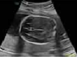
Background: Cavum septum pellucidum (CSP) is a critical anatomical land mark of normal midline development of brain and its absent is associated with brain anomalies. On the other hand, the size of CSP differs among different populations. The aim of this study was the evaluation of the size (width) of CSP at various gestation age (18 to 40 weeks’ pregnancy) in Isfahan, Iran population. Materials and Methods: This cross-sectional study was performed at Isfahan hospitals (Al-Zahra and Shahid Beheshti) from December 2022 until December 2023. The CSP was measured in 1000 normal fetuses at trans-ventricular plane by trans-abdominal ultrasound by two expert operators. In these fetuses the HC and BPD were measured. Regression analysis revealed significant relationship between CSP width and gestational age, also between CSP width and HC and BPD. Results: The mean CSP width was 5.3 ± 2.08 mm, ranging from 2.15 mm to 8.85 mm. CSP width increased progressively from 18 to 33 weeks, plateaued at 33–37 weeks, and slightly declined near term. Strong correlations were found between CSP width and head circumference (HC) (r=0.644, P<0.01) and biparietal diameter (BPD) (r=0.631, P<0.01). The study defined normative fetal CSP development values using percentile distributions with a 5th percentile length-to-width ratio of 1.06. Conclusion: The present study provides normative data for fetal CSP width and useful information about the development of the CSP based on gestational age.
References
Tubbs, R.S., et al., Cavum velum interpositum, cavum septum pellucidum, and cavum vergae: a review. Child's Nervous System, 2011. 27: p. 1927-1930
https://doi.org/10.1007/s00381-011-1457-2
Callen, P.W., et al., Columns of the fornix, not to be mistaken for the cavum septi pellucidi on prenatal sonography. Journal of Ultrasound in Medicine, 2008. 27(1): p. 25-31
https://doi.org/10.7863/jum.2008.27.1.25
Woodward, P.J., Diagnostic Imaging: Obstetrics: Diagnostic Imaging: Obstetrics E-Book. 2021: Elsevier Health Sciences.
Jou, H.J., et al., Ultrasound measurement of the fetal cavum septi pellucidi. Ultrasound in Obstetrics and Gynecology: The Official Journal of the International Society of Ultrasound in Obstetrics and Gynecology, 1998. 12(6): p. 419-421
https://doi.org/10.1046/j.1469-0705.1998.12060419.x
Paladini, D., et al., Sonographic examination of the fetal central nervous system: guidelines for performing the 'basic examination'and the 'fetal neurosonogram'. Ultrasound in Obstetrics & Gynecology, 2007. 29(1): p. 109-116
https://doi.org/10.1002/uog.3909
Siala, S., et al., Imaging of the septum pellucidum: normal, variants and pathology. The British Journal of Radiology, 2023. 96: p. 20221058
https://doi.org/10.1259/bjr.20221058
Saleh, K., E. Al-Ajmi, and A. Al Futaisi, Absent Septum Pellucidum: Search for other anomalies. Sultan Qaboos University Medical Journal, 2023. 23(3): p. 423
https://doi.org/10.18295/squmj.5.2023.024
Siriwardana, S.R. and C.K. Pathirage, Radiological Anatomy, Variations, and Clinical Significance of Septum Pellucidum: A Mini-Review. 2023
https://doi.org/10.20944/preprints202312.1486.v1
Ward, A., A. Monteagudo, and S.f.M.-F. Medicine, Absent cavum septi pellucidi. American Journal of Obstetrics and Gynecology, 2020. 223(6): p. B23-B26
https://doi.org/10.1016/j.ajog.2020.08.180
Bardini, R., Isolated, Absent Cavum Septum Pellucidum: A Single Center's Outcomes and Review of the Literature. 2021
Dremmen, M., et al., Cavum septum pellucidum in the general pediatric population and its relation to surrounding brain structure volumes, cognitive function, and emotional or behavioral problems. American Journal of Neuroradiology, 2019. 40(2): p. 340-346
https://doi.org/10.3174/ajnr.A5939
Falco, P., et al., Transabdominal sonography of the cavum septum pellucidum in normal fetuses in the second and third trimesters of pregnancy. Ultrasound Obstet Gynecol, 2000. 16(6): p. 549-53.
https://doi.org/10.1046/j.1469-0705.2000.00244.x
Rakic, P. and P.I. Yakovlev, Development of the corpus callosum and cavum septi in man. Journal of Comparative Neurology, 1968. 132(1): p. 45-72
https://doi.org/10.1002/cne.901320103
Arisoy, R., et al., Cavum septum pellucidum nomogram during the second trimester of pregnancy. Journal of Obstetrics and Gynaecology, 2022. 42(7): p. 2931-2934
https://doi.org/10.1080/01443615.2022.2114323
Serhatlioglu, S., et al., Sonographic measurement of the fetal cerebellum, cisterna magna, and cavum septum pellucidum in normal fetuses in the second and third trimesters of pregnancy. Journal of clinical ultrasound, 2003. 31(4): p. 194-200
https://doi.org/10.1002/jcu.10163
Tao, G., et al., Sonographic appearance of the cavum septum pellucidum et vergae in normal fetuses in the second and third trimesters of pregnancy. Journal of Clinical Ultrasound, 2013. 41(9): p. 525-531
https://doi.org/10.1002/jcu.22084
Dahmardeh, H., et al., Evaluating the Size Range of Cavum Septum Pellucidum in the Second and the Third Trimester of Pregnancy in Women Referred to Ali Ebn-e Abi Taleb Hospital of Zahedan. Zahedan Journal of Research in Medical Sciences, 2023. 25(1)
https://doi.org/10.5812/zjrms-119002
Karl, K., et al., Cavum septi pellucidi (CSP) ratio: a marker for partial agenesis of the fetal corpus callosum. Ultrasound in Obstetrics & Gynecology, 2017. 50(3): p. 336-341
Published
How to Cite
Issue
Section
License
Copyright (c) 2025 Galen Medical Journal

This work is licensed under a Creative Commons Attribution 4.0 International License.


