
Evaluation of Micronucleus Count in Oral Squamous Cell Carcinoma (OSCC) Newly Diagnosed Patients Compared to Previously Treated Ones: A Cytologic Study
DOI:
https://doi.org/10.31661/gmj.vi.3968Keywords:
Mouth Mucosa; Micronucleus Assays; Oral Squamous Cell Carcinoma; Cytology; ScreeningAbstract
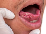
Background: Early detection and monitoring of genomic damages are vital for improving therapeutic outcomes. Quantification of micronuclei in exfoliated buccal mucosa cells has emerged as a reliable biomarker for assessing genomic alterations and cytogenetic damage in precancerous and cancerous conditions. Materials and Methods: This study evaluated exfoliated buccal cells from two groups of OSCC patients: seventeen newly diagnosed individuals who hadn’t yet undergone OSCC treatments and seventeen patients assessed at least six months after treatments. Micronuclei were identified and quantified in the cytology samples, and statistical analyses including the T-test, Mann–Whitney U, Kruskal–Wallis, and Spearman’s correlation tests were applied at a significance threshold of P<0.05 to compare modalities between groups. Results: The newly diagnosed group exhibited a mean micronucleus frequency of 0.028±0.013 per 10³ cell, whereas the treated group demonstrated a significantly lower mean frequency of 0.016±0.020 per 10³ cell (p=0.03). Further stratification of treated patients by intervention type (surgery alone, surgery combined with radiotherapy, and surgery followed by radiotherapy and chemotherapy) yielded mean counts of 0.006±0.003 per 10³ cell, 0.014±0.010 per 10³ cell, and 0.026±0.025 per 10³ cell, respectively. These variations did not reach statistical significance (p=0.29). Conclusion: The findings show that treatment reduces cytogenetic damage, as reflected by diminished micronucleus formation. Consequently, micronucleus assessment in buccal mucosa cells may serve as a noninvasive, cost-effective tool for monitoring therapeutic efficacy and predicting the recovery process in OSCC patients.
References
Proia NK, Paszkiewicz GM, Nasca MA, Franke GE, Pauly JL. Smoking and smokeless tobacco associated human buccal cell mutations and their association with oral cancer a review. Cancer Epidemiol Biomarkers Prev. 2006; 15:1061 77.
https://doi.org/10.1158/1055-9965.EPI-05-0983
PMid:16775162
Palve DH, Tupkari JV. Clinicopathological correlation of micronuclei in oral squamous cell carcinoma by exfoliative cytology. J Oral Maxillofac Pathol. 2008; 12:2 7.
https://doi.org/10.4103/0973-029X.42189
Chaudhary M, Venkatapathy R, Oza N, Prashad KV, Malik S. Evaluation of Micronuclei in Oral Squamous Cell Carcinoma: A Cytological Study. Int J Oral Care Res. 2017; 5(1):48.
https://doi.org/10.5005/jp-journals-10051-0074
Tolbert PE, Shy CM, Allen JW. Micronuclei and other nuclear anomalies in buccal smears: Methods development. Mutat Res. 1992; 271:69-77.
https://doi.org/10.1016/0165-1161(92)90033-I
PMid:1371831
Stich HF, Rosin MP, Vallejera MO. Reduction with vitamin A and betacarotene administration of proportion of micronucleated buccal mucosal cells in Asian betal nut and tobacco chewers. Lancet. 1984; 1:12046
https://doi.org/10.1016/S0140-6736(84)91692-1
PMid:6144923
Jalayer Naderi N, Pour Pasha M. Comparison of cytotoxic effect of cigarette and waterpipe smoking on human buccal mucosa. Int J Prev Med. 2017; 8:98.
https://doi.org/10.4103/ijpvm.IJPVM_62_17
PMid:29291040 PMCid:PMC5738785
Farhadi S, Jahanbani J, Sadri D, Moridani SG. The micronucleus assessment of buccal mucosa: A noninvasive method in screening of smokers potentially exposed to oral cancer. IJBPAS. 2015; 4(12): 68166825.
Nersesyan AK, Vardazaryan NS, Gevorgyan AL, Arutyunyan RM. Micronucleus level in exfoliated buccal mucosa cells of cancer patients. Arch Oncol. 2002; 10:35 6.
https://doi.org/10.2298/AOO0201035N
Khanna S, Purwar A, Singh NN, Sreedhar G, Singh S, Bhalla S. Cytogenetic biomonitoring of premalignant and malignant oral lesions by micronuclei assessment: A screening evaluation. European Journal of General Dentistry. 2014; 3(01):4652.
https://doi.org/10.4103/2278-9626.126211
Sivasankari P, Kaur S, Reddy KS, Vivekanandam S, Rao RK. Micronucleus index: An early diagnosis in oral carcinoma. J Anat Soc India. 2008; 57:8‑13.
Jalili S, Naderi NJ. Comparison of repair index in cigarette and waterpipe smokers: A biomonitoring assessment using human exfoliated buccal mucosa cells. International Journal of Preventive Medicine. 2022; 13:27.
https://doi.org/10.4103/ijpvm.IJPVM_10_20
PMid:35392320 PMCid:PMC8980829
Kumar V, Rao NN, Nair NS. Micronuclei in oral squamous cell carcinoma: A marker of genotoxic damage. Indian J Dent Res. 2000; 11:1016.
deCarvalho MB, Ramirez A, Gattás GJ, Guedes AL, Amar A, Rapoport A, et al. Relationship between the outcome and the frequency of micronuclei in cells of patients with oral and oropharyngeal carcinoma. Rev Assoc Med Bras. 2002; 48:31722.
https://doi.org/10.1590/S0104-42302002000400037
PMid:12563459
Tak A, Metgud R, Astekar M, Tak M. Micronuclei and other nuclear anomalies in normal human buccal mucosa cells of oral cancer patients undergoing radiotherapy: a field effect. Biotech Histochem. 2014; 89(6):4649.
https://doi.org/10.3109/10520295.2014.904925
PMid:24799093
Minicucci EM, Kowalski LP, Maia MA, Pereira A, Ribeiro LR, de Camargo JL, Salvadori DM. Cytogenetic damage in circulating lymphocytes and buccal mucosa cells of headandneck cancer patients undergoing radiotherapy. J Radiat Res. 2005; 46(2):13542.
https://doi.org/10.1269/jrr.46.135
PMid:15988130
Sangle VA, Bijjaragi S, Shah N, Kangane S, Ghule HM, Rani SA. Comparative study of frequency of micronuclei in normal, potentially malignant diseases and oral squamous cell carcinoma. J Nat Sc Biol Med. 2016; 7:338.
https://doi.org/10.4103/0976-9668.175049
PMid:27003966 PMCid:PMC4780163
Kiran K, Agarwal P, Kumar S, Jain K. Micronuclei as a predictor for oral carcinogenesis. J Cytol. 2018; 35:2336.
https://doi.org/10.4103/JOC.JOC_141_17
PMid:30498296 PMCid:PMC6210814
Driessens G, Harsan L, Robaye B, Waroquier D, Browaeys P, Giannakopoulos X, Velu T, Bruyns C. Micronuclei to detect in vivo chemotherapy damage in a p53 mutated solid tumour. Br J Cancer. 2003; 89(4):7279.
https://doi.org/10.1038/sj.bjc.6601163
PMid:12915886 PMCid:PMC2376913
Published
How to Cite
Issue
Section
License
Copyright (c) 2025 Galen Medical Journal

This work is licensed under a Creative Commons Attribution 4.0 International License.


