
Comparison of Energy and Frequency of Nanosecond Q-switched ND-YAG Laser and Chlorhexidine on Reducing the Number of Oral Streptococcus Mutans Isolates: In Vitro Study
DOI:
https://doi.org/10.31661/gmj.vi.3973Keywords:
Streptococcus Mutans; Chlorhexidine; Nd:YAG NanosecondAbstract
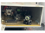
Background: Streptococcus mutans is a facultative anaerobic coccus that is a part of the oral flora of humans. chlorhexidine also has some side effects. Today, Nd: YAG lasers have become very popular in dentistry and are used for various types of treatment. Therefore, this study aims to compare the effects of Chlorhexidine 2% and nanosecond (Nd: YAG) laser in reducing the number of oral Streptococcus mutans bacteria in the oral cavity. Materials and Methods: An in vitro experimental design was conducted using S. mutans ATCC 35668 cultured on Mueller-Hinton agar. Bacterial suspensions were standardized to a half-McFarland turbidity and distributed in 96-well plates. The antimicrobial activity of 2% CHX was assessed via the well-diffusion method. Laser irradiation was applied at varying energies (10, 20, 30 mJ) and frequencies (5, 10 Hz). Bacterial counts and inhibition zone diameters were measured, and data were analyzed using two-way ANOVA followed by Tukey post hoc tests (α=0.05). Results: Bacterial counts decreased with increasing laser energy and frequency, with complete elimination observed at 30 mJ (both frequencies) and at 20 mJ/10 Hz. Inhibition zone diameters were largest at 30 mJ/10 Hz (8.6 ± 0.39 mm), while 2% CHX produced a slightly larger, but not statistically significant, inhibition zone (13 ± 2.9 mm vs. 11.5 ± 14.4 mm; P>.05). Both laser parameters and CHX significantly reduced S. mutans populations, demonstrating comparable antimicrobial efficacy at higher laser energies. Conclusion: High-energy 1064 nm Q-switched Nd:YAG laser therapy effectively reduces S. mutans counts in vitro, with results comparable to 2% CHX. Laser application may serve as an alternative or adjunctive strategy for microbial control in dental treatments, particularly in cases where chemical antiseptics are limited or contraindicated.
References
World Health Organization. Global tuberculosis report 2023 [Internet]. Geneva: World Health Organization; 2023. Available from: http://www.who.int/tb/en
Moaddab SR, Amini K, Haki K. Determining the drug susceptibility of Mycobacterium tuberculosis strains to pyrazinamide. Med J Tabriz Univ Med Sci. 2015;37:56-62.
Alavi SM, Enayatrad M, Cheraghian B, Amoori N. Incidence trend analysis of tuberculosis in Khuzestan Province, southwest of Iran: 2010-2019. Glob Epidemiol. 2023;6:100118.
https://doi.org/10.1016/j.gloepi.2023.100118
PMid:37637715 PMCid:PMC10445994
Kiani B, Raouf Rahmati A, Bergquist R, Hashtarkhani S, Firouraghi N, Bagheri N, Mohammadi A. Spatiotemporal epidemiology of the tuberculosis incidence rate in Iran, 2008-2018. BMC Public Health. 2021;21:1-20.
https://doi.org/10.1186/s12889-021-11157-1
PMid:34098917 PMCid:PMC8186231
Kim S, Hesseling AC, Wu X, Hughes MD, Shah NS, Gaikwad S, ACTG A5300/IMPAACT 2003 PHOENIx Feasibility Study Team. Factors associated with prevalent Mycobacterium tuberculosis infection and disease among adolescents and adults exposed to rifampinresistant tuberculosis in the household. PLoS One. 2021;18:e0283290.
https://doi.org/10.1371/journal.pone.0283290
PMid:36930628 PMCid:PMC10022776
Lake MA, Adams KN, Nie F, Fowler E, Verma AK, Dei S, Ramakrishnan L. The human proton pump inhibitors inhibit Mycobacterium tuberculosis rifampicin efflux and macrophageinduced rifampicin tolerance. Proc Natl Acad Sci U S A. 2023;120:e2215512120.
https://doi.org/10.1073/pnas.2215512120
PMid:36763530 PMCid:PMC7614234
Kaya H, Ersoy L, Ülger M, Bozok T, Aslan G. Investigation of efflux pump genes in isoniazidresistant Mycobacterium tuberculosis isolates. Indian J Med Microbiol. 2023;46:100428.
https://doi.org/10.1016/j.ijmmb.2023.100428
PMid:37945121
Desale VJ, Mali SN, Thorat BR, Yamgar RS, Dharanguttikar SV, Dharanguttikar VR, Cruz JN. Synthesis, characterization, ADMETSAR prediction, DPPH assay, and antiMycobacterium study of 4[(substituted benzyl)amino] benzo hydrazides and hydrazones as AcylCoA carboxylase AccD5 inhibitors. Curr Comput Aided Drug Des. 2023;19:300-312.
https://doi.org/10.2174/1573409919666221227091735
PMid:36578253
Rodrigues L, Cravo P, Viveiros M. Efflux pump inhibitors as a promising adjunct therapy against drugresistant tuberculosis: a new strategy to revisit mycobacterial targets and repurpose old drugs. Expert Rev Anti Infect Ther. 2020;18:741-757.
https://doi.org/10.1080/14787210.2020.1760845
PMid:32434397
Pitaloka DAE, Arfan A, Ramadhan DSF, Chaidir L. Insights into the molecular mechanism of pyrazinamide binding to mutated pyrazinamidase linked to the pncA gene in clinical isolates of Mycobacterium tuberculosis. J Biomol Struct Dyn. 2024;42:759-765.
https://doi.org/10.1080/07391102.2023.2195002
PMid:37096659
Modlin SJ, Mansjö M, Werngren J, Ejike CM, Hoffner SE, Valafar F. Pyrazinamideresistant tuberculosis is obscured from common targeted molecular diagnostics. Drug Resist Updat. 2023;68:100959.
https://doi.org/10.1016/j.drup.2023.100959
PMid:37043916 PMCid:PMC10317212
Alshabrmi FM, Alatawi EA. Deciphering the mechanism of resistance by novel double mutations in pncA in Mycobacterium tuberculosis using protein structural graphs and bioinformatic approaches. Comput Biol Med. 2023;154:106599.
https://doi.org/10.1016/j.compbiomed.2023.106599
PMid:36731361
van der Heijden YF, Maruri F, Blackman A, Morrison R, Guo Y, Sterling TR. Mycobacterium tuberculosis gene expression associated with fluoroquinolone resistance and efflux pump inhibition. J Infect Dis. 2023;228:469-478.
https://doi.org/10.1093/infdis/jiad112
PMid:37079382 PMCid:PMC10428193
Jawetz E, Melnick JL, Adelberg EA. Medical microbiology. 24th ed San Francisco: McGrawHill; 2007.
National Collaborating Centre for Chronic Conditions (Great Britain). Tuberculosis: clinical diagnosis and management of tuberculosis, and measures for its prevention and control. London: Royal College of Physicians; 2006.
Hashemzadeh M, Asareh Zadegan Dezfuli A, Moinikhah M, Yousefi F. Phenotypic and genotypic resistance to pyrazinamide in multidrugresistant Mycobacterium tuberculosis isolates in Khuzestan Province (2017-2022). Jundishapur Sci Med J. 2024;23:44-60. doi:10.32592/JSMJ.23.1.44.
https://doi.org/10.32592/JSMJ.23.1.44
Hashemzadeh M, Asareh Zadegan Dezfuli A, Khosravi A, Ahmad Khosravi N, Saki M. Antibiotic resistance and genomic characterization of Mycobacterium abscessus complex isolates from pulmonary tuberculosis patients in Iran: a multicenter study (2010-2021). Acta Microbiol Immunol Hung. 2024;71:82-88.
https://doi.org/10.1556/030.2024.02169
PMid:38285119
Zhang Y, Zhang J, Cui P, Zhang Y, Zhang W. Identification of novel efflux proteins Rv0191, Rv3756c, Rv3008, and Rv1667c involved in pyrazinamide resistance in Mycobacterium tuberculosis. Antimicrob Agents Chemother. 2017;61:e0112816.
https://doi.org/10.1128/AAC.00940-17
PMid:28584158 PMCid:PMC5527661
GrzechLeśniak Z, Szwach J, Lelonkiewicz M, Migas K, Pyrkosz J, Szwajkowski M, Kosidło P, Pajączkowska M, Wiench R, Matys J, Nowicka J. Effect of Nd: YAG Laser Irradiation on the Growth of Oral Biofilm. Microorganisms. 2024 Nov 4;12(11):2231.
https://doi.org/10.3390/microorganisms12112231
PMid:39597620 PMCid:PMC11596257
Namour M, Verspecht T, El Mobadder M, Teughels W, Peremans A, Nammour S, Rompen E. QSwitch Nd: YAG laserassisted elimination of multispecies biofilm on titanium surfaces. Materials. 2020 Mar 29;13(7):1573.
https://doi.org/10.3390/ma13071573
PMid:32235332 PMCid:PMC7177273
Golob Deeb J, Reddy N, Kitten T, Carrico CK, GrzechLeśniak K. Viability of bacteria associated with root caries after Nd: YAG laser application in combination with various antimicrobial agents: An in vitro study. Dental and Medical Problems. 2023;60(4):64955.
https://doi.org/10.17219/dmp/171690
PMid:37982598
Anwer AG, Markaryan KW. Bactericidal effect of Qswitched Nd: YAG laser (1064 nm) on Streptococcus mutans from carious teeth, in vitro study. Iraqi Journal of Laser. 2009 Dec 10;8(B):516.
Namour M, El Mobadder M, Mulongo B, Fagnart O, Harb A, Peremans A, Verspecht T, Teughels W, Nammour S, Rompen E. Assessment of disinfection potential of QSwitch Nd: YAG laser on contaminated titanium implant surfaces. Materials. 2021 Oct 14;14(20):6078.
https://doi.org/10.3390/ma14206078
PMid:34683666 PMCid:PMC8537820
Ibrahim GA, Al Sarraf IA, Sadik BH, Noori ST, Mohammed AA. Effect of Nd. YAG laser on microorganisms causing dental caries: an in vitro study Front Biomed Technol; 2025.
Published
How to Cite
Issue
Section
License
Copyright (c) 2025 Galen Medical Journal

This work is licensed under a Creative Commons Attribution 4.0 International License.


