
Prevalence and Distribution of Sagittal and Vertical Subtypes in Skeletal Class III Malocclusion: A Retrospective Study across Age and Sex Groups
DOI:
https://doi.org/10.31661/gmj.v14iSP1.4020Keywords:
Sagittal Subtypes; Vertical Subtypes; Skeletal Class III MalocclusionAbstract
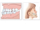
Background: Skeletal Class III malocclusion causing mandibular prognathism, maxillary retrognathism, or a combination of both, has widely varying prevalence by ethnicity. The objective of this research is to investigate the prevalence of contributing factors in its development across different age and sex groups of Iranians. Materials and Methods: In this study, 233 lateral cephalograms of patients with skeletal Class III malocclusion who had referred our orthodontics center from 2015 to 2024. In the sagittal dimension, the samples were categorized into four groups: retrognathic maxilla, prognathic mandible, combination, and normal. The prevalence of each condition was analyzed across groups. In case of discrepancies between Steiner and McNamara analyses, final sagittal diagnoses were manually determined. Results: This study examined 233 lateral cephalograms of individuals with skeletal Class III malocclusion, including 101 males (43.3%) and 132 females (56.7%). There was a significant association between the final sagittal relationship diagnosis and the combined age-sex groups (P=0.010), indicating that the distribution of sagittal patterns varies across age and sex subgroups. Mandibular prognathism was the most prevalent condition in all groups except for females aged 7–11, where mandibular prognathism and maxillary retrognathism were equally common. In the vertical dimension, 51.9% of cases had normal facial height. No significant correlation was found between vertical dimension and age or sex (P=0.479). The strongest positive correlation was observed between Sum.o.p and SN.GoMe, as well as Wits and A. NPPD, while the strongest negative correlations involved Jarabak.index with Sum.o.p and SN.GoMe. Conclusion: The results indicated that mandibular prognathism is the predominant cause of skeletal Class III malocclusion, except in females aged 7–11, where maxillary retrognathism is also prevalent. No discernible pattern was detected in vertical classification among age and sex groups. The research shows the significance of employing various diagnostic methods for a thorough assessment.
References
Kozanecka A, Sarul M, Kawala B, AntoszewskaSmith J. Objectification of Orthodontic Treatment Needs: Does the Classification of Malocclusions or a History of Orthodontic Treatment Matter? Adv Clin Exp Med. 2016;25(6):130312.
https://doi.org/10.17219/acem/62828
PMid:28028986
Ellis E, McNamara JA. Components of adult Class III malocclusion. J Oral Maxillofac Surg. 1984;42(5):295305.
https://doi.org/10.1016/0278-2391(84)90109-5
PMid:6585502
Sanborn RT. Differences Between the Facial Skeletal Patterns Of Class III Malocclusion and Normal Occlusion*. The Angle Orthodontist. 1955;25(4):20822.
Azamian Z, Shirban F. Treatment Options for Class III Malocclusion in Growing Patients with Emphasis on Maxillary Protraction. Scientifica. 2016;2016:8105163.
https://doi.org/10.1155/2016/8105163
PMid:27144056 PMCid:PMC4842067
William Proffit HF, Brent Larson, David Sarver. Contemporary Orthodontics. 6th: Edition ed; 2018.
StornioloSouza JM, Seminario MP, PinzanVercelino CRM, Pinzan A, Janson G. McNamara analysis cephalometric parameters in WhiteBrazilians, Japanese and JapaneseBrazilians with normal occlusion. Dental Press J Orthod. 2021;26(1):e2119133.
https://doi.org/10.1590/2177-6709.26.1.e2119133.oar
PMid:33759961 PMCid:PMC8018756
Esmaeili S, Mohammadi NM, Khosravani S, Eslamian L, Motamedian SR. Evaluation of facial profile characteristics of aesthetically pleasing Iranian faces. J World Fed Orthod. 2023;12(2):7689.
https://doi.org/10.1016/j.ejwf.2023.02.001
PMid:36906490
Baik HS, Han HK, Kim DJ, Proffit WR. Cephalometric characteristics of Korean Class III surgical patients and their relationship to plans for surgical treatment. Int J Adult Orthodon Orthognath Surg. 2000;15(2):11928.
McNamara JA. A method of cephalometric evaluation. American Journal of Orthodontics. 1984;86(6):44969.
https://doi.org/10.1016/S0002-9416(84)90352-X
PMid:6594933
Mouakeh M. Cephalometric evaluation of craniofacial pattern of syrian children with class III malocclusion. American Journal of Orthodontics and Dentofacial Orthopedics. 2001;119(6):6409.
https://doi.org/10.1067/mod.2001.112671
PMid:11395709
Staudt CB, Kiliaridis S. Different skeletal types underlying Class III malocclusion in a random population. Am J Orthod Dentofacial Orthop. 2009;136(5):71521.
https://doi.org/10.1016/j.ajodo.2007.10.061
PMid:19892290
Spalj S, Mestrovic S, Lapter Varga M, Slaj M. Skeletal components of class III malocclusions and compensation mechanisms. J Oral Rehabil. 2008;35(8):62937.
https://doi.org/10.1111/j.1365-2842.2008.01869.x
PMid:18699972
Paixão MB, Sobral MC, Vogel CJ, Araujo TM. Comparative study between manual and digital cephalometric tracing using Dolphin Imaging software with lateral radiographs. Dental Press Journal of Orthodontics. 2010;15:12330.
https://doi.org/10.1590/S2176-94512010000600016
Koodaryan R, Rafighi A, Hafezeqoran A. Components of Adult Class III Malocclusion in an Iranian Population. J Dent Res Dent Clin Dent Prospects. 2009;3(1):203.
K T, Naveen K, Prasanna Arvind TR. PREVALENCE OF CLASS III SKELETAL PATTERN AMONG PATIENTS SEEKING ORTHODONTIC TREATMENT. Journal of Pharmaceutical Negative Results. 2022:273641.
Li C, Cai Y, Chen S, Chen F. Classification and characterization of class III malocclusion in Chinese individuals. Head Face Med. 2016;12(1):31.
https://doi.org/10.1186/s13005-016-0127-8
PMid:27821165 PMCid:PMC5100215
Ramezanzadeh B, Pousti M, Bagheri M. Cephalometric Evaluation of Dentofacial Features of Class III Malocclusion in Adults of Mashhad, Iran. J Dent Res Dent Clin Dent Prospects. 2007;1(3):12530.
Toms AP. Class III malocclusion: a cephalometric study of Saudi Arabians. Br J Orthod. 1989;16(3):2016.
https://doi.org/10.1179/bjo.16.3.201
PMid:2765469
Kao CT, Chen FM, Lin TY, Huang TH. The craniofacial morphologic structures of the adult with Class III malocclusion. Int J Adult Orthodon Orthognath Surg. 1995;10(4):28593.
Ishii N, Deguchi T, Hunt NP. Craniofacial differences between Japanese and British Caucasian females with a skeletal Class III malocclusion. Eur J Orthod. 2002;24(5):4939.
https://doi.org/10.1093/ejo/24.5.493
PMid:12407945
Aljabaa A, Aldrees A. ANB, Wits And Molar Relationship, Do They Correlate In Orthodontic Patients? Dentistry. 2014;4:1.
https://doi.org/10.4172/2161-1122.1000240
Jabbar A, Mahmood A. Correlation of overjet, ANB and wits appraisal for assessment of sagittal skeletal relationship. Pakistan Orthodontic Journal. 2012;4(1):1723.
Ardani I, Willyanti I, Narmada IB. Correlation between vertical components and skeletal Class II malocclusion in ethnic Javanese. Clin Cosmet Investig Dent. 2018;10:297302.
https://doi.org/10.2147/CCIDE.S188414
PMid:30588125 PMCid:PMC6304072
Jeong S, Kim S, Lim SH, Yu SK. A study of correlations between cephalometric measurements in Koreans with normal occlusion by network analysis. Scientific Reports. 2024;14(1):9660.
https://doi.org/10.1038/s41598-024-60410-1
PMid:38671196 PMCid:PMC11053105
Published
How to Cite
Issue
Section
License
Copyright (c) 2025 Galen Medical Journal

This work is licensed under a Creative Commons Attribution 4.0 International License.


