
Evaluation of Morphologic Dimensions of Humulus Appendix of Pterygoid Plate using Cone Beam Computed Tomography (CBCT) in Ahvazian patients, Iran
DOI:
https://doi.org/10.31661/gmj.vi.3901Keywords:
Cone Beam Computed Tomography; Inclination; Axial; Coronal; Length; Width; Humulus of Pterygoid AppendageAbstract
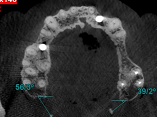
Background: Today, CBCT has found a special place in dentistry due to the high quality and accuracy of images and providing information, and its use is increasing. With its help, we can examine many parts of the anatomy that are difficult to evaluate. The purpose of this study is to investigate the morphological dimensions of the humulus of the pterygoid appendage using cone beam computed tomography (CBCT). Materials and Methods: In this retrospective study, the statistical population was the imaging reccords of patients who referred to the radiology department of Ahvaz Dental School for CBCT of the upper jaw, whose values were stored in the NNT software. The size of the humulus (length and width) and its slope in the coronal and axial sections of the images were evaluated by two oral and maxillofacial radiologists. The results were analyzed using SPSS software version 22. Results: Eighty pterygoid hamuli from 38 males and 42 females (age 26–64 years, mean 43.18 ± 11.57) were analyzed. No significant gender differences were observed in length (p = 0.096), width (p = 0.300), axial angle (p = 0.067), or coronal angle (p = 0.102). Age-related comparisons revealed significant variation: hamular length and width increased in the 31–40 and >51 year groups versus <30 years (p = 0.022–0.031), axial angle was higher in 31–40 and >51 year groups (p = 0.003–0.006), and coronal angle increased in 31–40 and >51 year groups (p = 0.047–0.049). Conclusion: These findings indicate gradual morphometric changes with age, independent of gender. The length of PH increases with age and then decreases. While the width increases with age. There was no significant difference between length and width measurements in men and women. These findings help to diagnose vague pains in the oropharynx region related to the altered morphology of PH.
References
Barchetta N F, Oliveira R L B de, Silveira V Á S et al. Clinical and morphofunctional aspects of pterygoid hamulus: literature review. Brazilian Dental Science. 2015; 18 (4): 5-11.
https://doi.org/10.14295/bds.2015.v18i4.1078
Rusu MC, Didilescu AC, Jianu AM, Păduraru D. 3D CBCT anatomy of the pterygopalatine fossa. Surgical and radiologic anatomy. 2013 Mar 1;35(2):14359.
https://doi.org/10.1007/s00276-012-1009-9
Iwanaga J, Kido J, Lipski M, Tomaszewska IM, Tomaszewski KA, Walocha JA, Oskouian RJ, Tubbs RS. Anatomical study of the palatine aponeurosis: application to posterior palatal seal of the complete maxillary denture. Surgical and Radiologic Anatomy. 2018 Feb;40(2):17983.
https://doi.org/10.1007/s00276-017-1911-2
PMid:28823003
White SC, Pharoah MJ. Oral RadiologyEBook. Principles and Interpretation: Elsevier Health Sciences; 2014.
Stratmann U, Mokrys K, Meyer U, Kleinheinz J, Joos U, Dirksen D, Bollmann F. Clinical anatomy and palpability of the inferior lateral pterygoid muscle. The Journal of prosthetic dentistry. 2000 May 1;83(5):54854.
https://doi.org/10.1016/S0022-3913(00)70013-8
PMid:10793387
Putz R, Kroyer A. Functional morphology of the pterygoid hamulus. Annals of anatomy= Anatomischer Anzeiger: official organ of the Anatomische Gesellschaft. 1999 Jan 1;181(1):858.
https://doi.org/10.1016/S0940-9602(99)80099-5
PMid:10081567
KrmpotićNemanić J, Vinter I, Marušić A. Relations of the pterygoid hamulus and hard palate in children and adults: anatomical implications for the function of the soft palate. Annals of AnatomyAnatomischer Anzeiger. 2006 Jan 3;188(1):6974.
https://doi.org/10.1016/j.aanat.2005.05.005
PMid:16447915
Abe M, Murakami G, Noguchi M, Kitamura S, Shimada K, Kohama GI. Variations in the tensor veli palatini muscle with special reference to its origin and insertion. The Cleft palatecraniofacial journal. 2004 Sep;41(5):47484.
https://doi.org/10.1597/02-049.1
PMid:15352865
Eyrich GK, Locher MC, Warnke T, Sailer HF. The pterygoid hamulus as a paininducing factor: A report of a case and a radiographic study. International journal of oral and maxillofacial surgery. 1997 Aug 1;26(4):2757.
https://doi.org/10.1016/S0901-5027(97)80867-3
PMid:9258718
Orhan K, Sakul BU, Oz U, Bilecenoglu B. Evaluation of the pterygoid hamulus morphology using cone beam computed tomography. Oral Surgery, Oral Medicine, Oral Pathology, Oral Radiology, and Endodontology. 2011 Aug 1;112(2):e4855.
https://doi.org/10.1016/j.tripleo.2011.02.038
PMid:21612951
Sasaki T, Imai Y, Fujibayashi T. A case of elongated pterygoid hamulus syndrome. Oral diseases. 2001 Mar;7(2):1313.
https://doi.org/10.1034/j.1601-0825.2001.70212.x
PMid:11355439
Arya S, Singhal P, Vengal M, Patil N, Bhateja S. Hamular bursitis: difficult to diagnose in orofacial pain. Pakistan Oral and Dental Journal. 2015;35(1): 3-6.
https://doi.org/10.5958/0976-156X.2014.00006.9
Cho JY, Cheon KY, Shin DW, Chun WB, Lee H. Pterygoid hamulus bursitis as a cause of craniofacial pain: a case report. Journal of the Korean Association of Oral and Maxillofacial Surgeons. 2013;39(3): 8 .
https://doi.org/10.5125/jkaoms.2013.39.3.134
PMid:24471031 PMCid:PMC3858168
Fu Y, Peng J, Chen W. The pterygoid hamulus syndrome with the main discomfort of pharynx. Lin chuang er bi yan hou ke za zhi= Journal of clinical otorhinolaryngology. 2004;18(3): 5 .
Kumar V, Ludlow J, Mol A, Cevidanes L. Comparison of conventional and cone beam CT synthesized cephalograms. Dentomaxillofacial Radiology. 2007; 36(5): 9.
https://doi.org/10.1259/dmfr/98032356
PMid:17586852
Robinson S, Suomalainen A, Kortesniemi M. mCT. Eur J Radiol. 2005;56:18591.
https://doi.org/10.1016/j.ejrad.2005.03.022
PMid:16233892
Arai Y, Tammisalo E, Iwai K, Hashimoto K, Shinoda K. Development of a compact computed tomographic apparatus for dental use. Dentomaxillofac Radiol. 1999;28(4):245-248.
https://doi.org/10.1038/sj/dmfr/4600448
PMid:10455389
Nickenig HJ, Eitner S. Reliability of implant placement after virtual planning of implant positions using cone beam CT data and surgical guide templates. J Craniomaxillofac Surg. 2007;35:207-211.
https://doi.org/10.1016/j.jcms.2007.02.004
PMid:17576068
Scarfe WC, Farman AG, Sukovic P. Clinical applications of conebeam computed tomography in dental practice. J Can Dent Assoc. 2006;71:7580.
Romoozi E, Razavi SH, Barouti P, Rahimi M. Investigating the morphologic indices of the hamulus pterygoid process using the CBCT technique. J Res Med Dent Sci. 2018 Mar 1;6:2404.
Mahdavi Z, Hafezi L. Assessment of the pterygoid hamulus dimensions and morphology in Iranian women using conebeam computed tomography. Journal of Dental Research, Dental Clinics, Dental Prospects. 2024 Dec 14;18(4):297.
https://doi.org/10.34172/joddd.40976
PMid:39895678 PMCid:PMC11786002
Khoubivand M, Ghaffari R, Anjomshoa M. Morphological evaluation of pterygoid hamulus through mprconebeam computed tomography images. Bull Env Pharmacol Life Sci. 2015 Jan 2;4:14450.
Sivadas KS, Ajila V, Hegde S, Jain Y. Evaluation of the morphology of pterygoid hamulus using cone beam computed tomography: A retrospective study. Journal of Oral Biology and Craniofacial Research. 2025 Nov 1;15(6):12549.
https://doi.org/10.1016/j.jobcr.2025.08.008
PMid:40837828 PMCid:PMC12361749
Mehra A, Karjodkar FR, Sansare K, Kapoor R, Tambawala S, Saxena VS. Assessment of the dimensions of the pterygoid hamulus for establishing ageand sexspecific reference standards using conebeam computed tomography. Imaging science in dentistry. 2021 Mar;51(1):49.
https://doi.org/10.5624/isd.20200184
PMid:33828961 PMCid:PMC8007398
KrmpotićNemanić J, Vinter I, Marusić A. Relations of the pterygoid hamulus and hard palate in children and adults: anatomical implications for the function of the soft palate. Ann Anat. 2006;188:69-74.
https://doi.org/10.1016/j.aanat.2005.05.005
PMid:16447915
Komarnitki I, MankowskaPliszka H, Ungier E, Dziedzic D, Grzegorczyk M, Tomczyk A,et al. Functional morphometry of the pterygoid hamulus A comparative study of modern and medieval populations. Anthropological Review. 2019;82(4):38995.
https://doi.org/10.2478/anre-2019-0029
Nerkar A, Gadgil R, Bhoosreddy A, Shah K, Varma S. Conebeam computed tomography study of morphometric evaluation of pterygoid hamulus. Int J Recent Adv Multidiscip Res. 2017;4:2678-2682.
Alalawi W, Kolarkodi SH. CBCTBased Morphometric Evaluation of Pterygoid Plates for Surgical Applications: A Retrospective Study in a Saudi Population Clinical article . J Int Dent Med Res. 2025; 18(3): 12681274.
Published
How to Cite
Issue
Section
License
Copyright (c) 2025 Galen Medical Journal

This work is licensed under a Creative Commons Attribution 4.0 International License.


